1FQG

MOLECULAR STRUCTURE OF THE ACYL-ENZYME INTERMEDIATE IN TEM-1 BETA-LACTAMASE


Experimental Data Snapshot

  • Method: X-RAY DIFFRACTION
  • Resolution: 1.70 Å
  • R-Value Work: 0.182 
  • R-Value Observed: 0.182 

wwPDB Validation   3D Report Full Report


This is version 1.4 of the entry. See complete history


Literature

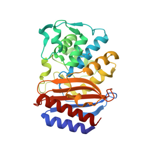
Molecular structure of the acyl-enzyme intermediate in beta-lactam hydrolysis at 1.7 A resolution.

Strynadka, N.C.Adachi, H.Jensen, S.E.Johns, K.Sielecki, A.Betzel, C.Sutoh, K.James, M.N.

(1992) Nature 359: 700-705

  • DOI: https://doi.org/10.1038/359700a0
  • Primary Citation of Related Structures:  
    1FQG

  • PubMed Abstract: 

    The X-ray crystal structure of the molecular complex of penicillin G with a deacylation-defective mutant of the RTEM-1 beta-lactamase from Escherichia coli shows how these antibiotics are recognized and destroyed. Penicillin G is covalently bound to Ser 70 0 gamma as an acyl-enzyme intermediate. The deduced catalytic mechanism uses Ser 70 0 gamma as the attacking nucleophile during acylation. Lys 73 N zeta acts as a general base in abstracting a proton from Ser 70 and transferring it to the thiazolidine ring nitrogen atom via Ser 130 0 gamma. Deacylation is accomplished by nucleophilic attack on the penicilloyl carbonyl carbon by a water molecule assisted by the general base, Glu 166.


  • Organizational Affiliation

    Department of Biochemistry, University of Alberta, Edmonton, Canada.


Macromolecules
Find similar proteins by:  (by identity cutoff)  |  3D Structure
Entity ID: 1
MoleculeChains Sequence LengthOrganismDetailsImage
TEM-1 BETA-LACTAMASE263Escherichia coliMutation(s): 1 
EC: 3.5.2.6
UniProt
Find proteins for P62593 (Escherichia coli)
Explore P62593 
Go to UniProtKB:  P62593
Entity Groups  
Sequence Clusters30% Identity50% Identity70% Identity90% Identity95% Identity100% Identity
UniProt GroupP62593
Sequence Annotations
Expand
  • Reference Sequence
Small Molecules
Ligands 1 Unique
IDChains Name / Formula / InChI Key2D Diagram3D Interactions
PNM
Query on PNM

Download Ideal Coordinates CCD File 
B [auth A]OPEN FORM - PENICILLIN G
C16 H20 N2 O4 S
OGFZUTGOGYUTKZ-KWCYVHTRSA-N
Experimental Data & Validation

Experimental Data

  • Method: X-RAY DIFFRACTION
  • Resolution: 1.70 Å
  • R-Value Work: 0.182 
  • R-Value Observed: 0.182 
  • Space Group: P 21 21 21
Unit Cell:
Length ( Å )Angle ( ˚ )
a = 62.2α = 90
b = 88.3β = 90
c = 41.6γ = 90
Software Package:
Software NamePurpose
MAR345data collection
ROTAVATAdata reduction
MLPHAREphasing
TNTrefinement
CCP4data scaling
ROTAVATAdata scaling

Structure Validation

View Full Validation Report


Entry History 

Deposition Data

Revision History  (Full details and data files)

  • Version 1.0: 2000-11-01
    Type: Initial release
  • Version 1.1: 2008-04-27
    Changes: Version format compliance
  • Version 1.2: 2011-07-13
    Changes: Version format compliance
  • Version 1.3: 2017-10-04
    Changes: Refinement description
  • Version 1.4: 2021-11-03
    Changes: Data collection, Database references, Derived calculations