1FQ5

X-ray structure of a cyclic statine inhibitor PD-129,541 bound to yeast proteinase A


Experimental Data Snapshot

  • Method: X-RAY DIFFRACTION
  • Resolution: 2.40 Å
  • R-Value Free: 0.280 
  • R-Value Observed: 0.200 

wwPDB Validation   3D Report Full Report


Ligand Structure Quality Assessment 


This is version 2.0 of the entry. See complete history


Literature

X-ray structures of five renin inhibitors bound to saccharopepsin: exploration of active-site specificity.

Cronin, N.B.Badasso, M.O.J Tickle, I.Dreyer, T.Hoover, D.J.Rosati, R.L.Humblet, C.C.Lunney, E.A.Cooper, J.B.

(2000) J Mol Biol 303: 745-760

  • DOI: https://doi.org/10.1006/jmbi.2000.4181
  • Primary Citation of Related Structures:  
    1FQ4, 1FQ5, 1FQ6, 1FQ7, 1FQ8

  • PubMed Abstract: 

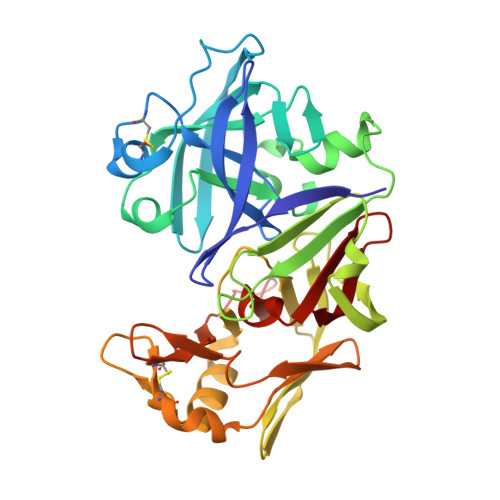
    Saccharopepsin is a vacuolar aspartic proteinase involved in activation of a number of hydrolases. The enzyme has great structural homology to mammalian aspartic proteinases including human renin and we have used it as a model system to study the binding of renin inhibitors by X-ray crystallography. Five medium-to-high resolution structures of saccharopepsin complexed with transition-state analogue renin inhibitors were determined. The structure of a cyclic peptide inhibitor (PD-129,541) complexed with the proteinase was solved to 2.5 A resolution. This inhibitor has low affinity for human renin yet binds very tightly to the yeast proteinase (K(i)=4 nM). The high affinity of this inhibitor can be attributed to its bulky cyclic moiety spanning P(2)-P(3)' and other residues that appear to optimally fit the binding sub-sites of the enzyme. Superposition of the saccharopepsin structure on that of renin showed that a movement of the loop 286-301 relative to renin facilitates tighter binding of this inhibitor to saccharopepsin. Our 2.8 A resolution structure of the complex with CP-108,420 shows that its benzimidazole P(3 )replacement retains one of the standard hydrogen bonds that normally involve the inhibitor's main-chain. This suggests a non-peptide lead in overcoming the problem of susceptible peptide bonds in the design of aspartic proteinase inhibitors. CP-72,647 which possesses a basic histidine residue at P(2), has a high affinity for renin (K(i)=5 nM) but proves to be a poor inhibitor for saccharopepsin (K(i)=3.7 microM). This may stem from the fact that the histidine residue would not bind favourably with the predominantly hydrophobic S(2) sub-site of saccharopepsin.


  • Organizational Affiliation

    Department of Crystallography, Birkbeck College, University of London, London, WC1E 7HX, UK.


Macromolecules
Find similar proteins by:  (by identity cutoff)  |  3D Structure
Entity ID: 1
MoleculeChains Sequence LengthOrganismDetailsImage
SACCHAROPEPSIN329Saccharomyces cerevisiaeMutation(s): 0 
EC: 3.4.23.25
Membrane Entity: Yes 
UniProt
Find proteins for P07267 (Saccharomyces cerevisiae (strain ATCC 204508 / S288c))
Explore P07267 
Go to UniProtKB:  P07267
Entity Groups  
Sequence Clusters30% Identity50% Identity70% Identity90% Identity95% Identity100% Identity
UniProt GroupP07267
Sequence Annotations
Expand
  • Reference Sequence
Oligosaccharides

Help

Entity ID: 2
MoleculeChains Length2D Diagram Glycosylation3D Interactions
beta-D-mannopyranose-(1-2)-alpha-D-mannopyranose-(1-3)-alpha-D-mannopyranose-(1-4)-2-acetamido-2-deoxy-beta-D-glucopyranose-(1-4)-2-acetamido-2-deoxy-beta-D-glucopyranose
B
5N-Glycosylation
Glycosylation Resources
GlyTouCan:  G28438DM
GlyCosmos:  G28438DM
GlyGen:  G28438DM
Small Molecules
Ligands 2 Unique
IDChains Name / Formula / InChI Key2D Diagram3D Interactions
0GM
Query on 0GM

Download Ideal Coordinates CCD File 
D [auth A]N-[(5S,9S,10S,13S)-9-hydroxy-5,10-bis(2-methylpropyl)-4,7,12,16-tetraoxo-3,6,11,17-tetraazabicyclo[17.3.1]tricosa-1(23),19,21-trien-13-yl]-3-(naphthalen-1-yl)-2-(naphthalen-1-ylmethyl)propanamide
C51 H61 N5 O6
KFCMUWOMPCWWCH-AXYJRABVSA-N
NAG
Query on NAG

Download Ideal Coordinates CCD File 
C [auth A]2-acetamido-2-deoxy-beta-D-glucopyranose
C8 H15 N O6
OVRNDRQMDRJTHS-FMDGEEDCSA-N
Binding Affinity Annotations 
IDSourceBinding Affinity
0GM Binding MOAD:  1FQ5 Ki: 4 (nM) from 1 assay(s)
Experimental Data & Validation

Experimental Data

  • Method: X-RAY DIFFRACTION
  • Resolution: 2.40 Å
  • R-Value Free: 0.280 
  • R-Value Observed: 0.200 
  • Space Group: P 32 2 1
Unit Cell:
Length ( Å )Angle ( ˚ )
a = 87.3α = 90
b = 87.3β = 90
c = 110.7γ = 120
Software Package:
Software NamePurpose
MOSFLMdata reduction
ROTAVATAdata reduction
AMoREphasing
RESTRAINrefinement
CCP4data scaling
ROTAVATAdata scaling

Structure Validation

View Full Validation Report


Ligand Structure Quality Assessment 


Entry History 

Deposition Data

Revision History  (Full details and data files)

  • Version 1.0: 2000-09-20
    Type: Initial release
  • Version 1.1: 2008-04-01
    Changes: Version format compliance
  • Version 1.2: 2011-07-13
    Changes: Atomic model, Database references, Derived calculations, Non-polymer description, Structure summary, Version format compliance
  • Version 1.3: 2016-12-14
    Changes: Structure summary
  • Version 1.4: 2017-10-04
    Changes: Refinement description
  • Version 2.0: 2020-07-29
    Type: Remediation
    Reason: Carbohydrate remediation
    Changes: Advisory, Atomic model, Data collection, Database references, Derived calculations, Non-polymer description, Structure summary