1FPZ

CRYSTAL STRUCTURE ANALYSIS OF KINASE ASSOCIATED PHOSPHATASE (KAP) WITH A SUBSTITUTION OF THE CATALYTIC SITE CYSTEINE (CYS140) TO A SERINE


Experimental Data Snapshot

  • Method: X-RAY DIFFRACTION
  • Resolution: 2.00 Å
  • R-Value Free: 0.253 
  • R-Value Work: 0.202 

wwPDB Validation   3D Report Full Report


This is version 1.4 of the entry. See complete history


Literature

Phosphoprotein-protein interactions revealed by the crystal structure of kinase-associated phosphatase in complex with phosphoCDK2.

Song, H.Hanlon, N.Brown, N.R.Noble, M.E.Johnson, L.N.Barford, D.

(2001) Mol Cell 7: 615-626

  • DOI: https://doi.org/10.1016/s1097-2765(01)00208-8
  • Primary Citation of Related Structures:  
    1FPZ, 1FQ1

  • PubMed Abstract: 

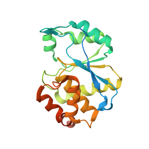
    The CDK-interacting protein phosphatase KAP dephosphorylates phosphoThr-160 (pThr-160) of the CDK2 activation segment, the site of regulatory phosphorylation that is essential for kinase activity. Here we describe the crystal structure of KAP in association with pThr-160-CDK2, representing an example of a protein phosphatase in complex with its intact protein substrate. The major protein interface between the two molecules is formed by the C-terminal lobe of CDK2 and the C-terminal helix of KAP, regions remote from the kinase-activation segment and the KAP catalytic site. The kinase-activation segment interacts with the catalytic site of KAP almost entirely via the phosphate group of pThr-160. This interaction requires that the activation segment is unfolded and drawn away from the kinase molecule, inducing a conformation of CDK2 similar to the activated state observed in the CDK2/cyclin A complex.


  • Organizational Affiliation

    Department of Biochemistry, University of Oxford, United Kingdom.


Macromolecules
Find similar proteins by:  (by identity cutoff)  |  3D Structure
Entity ID: 1
MoleculeChains Sequence LengthOrganismDetailsImage
CYCLIN-DEPENDENT KINASE INHIBITOR 3
A, B, C, D, E
A, B, C, D, E, F
212Homo sapiensMutation(s): 1 
EC: 3.1.3.48
UniProt & NIH Common Fund Data Resources
Find proteins for Q16667 (Homo sapiens)
Explore Q16667 
Go to UniProtKB:  Q16667
PHAROS:  Q16667
GTEx:  ENSG00000100526 
Entity Groups  
Sequence Clusters30% Identity50% Identity70% Identity90% Identity95% Identity100% Identity
UniProt GroupQ16667
Sequence Annotations
Expand
  • Reference Sequence
Experimental Data & Validation

Experimental Data

  • Method: X-RAY DIFFRACTION
  • Resolution: 2.00 Å
  • R-Value Free: 0.253 
  • R-Value Work: 0.202 
  • Space Group: P 65
Unit Cell:
Length ( Å )Angle ( ˚ )
a = 131.93α = 90
b = 131.93β = 90
c = 140.24γ = 120
Software Package:
Software NamePurpose
AMoREphasing
CNSrefinement
MOSFLMdata reduction
CCP4data scaling

Structure Validation

View Full Validation Report


Entry History 

Deposition Data

Revision History  (Full details and data files)

  • Version 1.0: 2001-05-09
    Type: Initial release
  • Version 1.1: 2008-04-27
    Changes: Version format compliance
  • Version 1.2: 2011-07-13
    Changes: Version format compliance
  • Version 1.3: 2018-03-14
    Changes: Database references
  • Version 1.4: 2024-02-07
    Changes: Data collection, Database references, Derived calculations