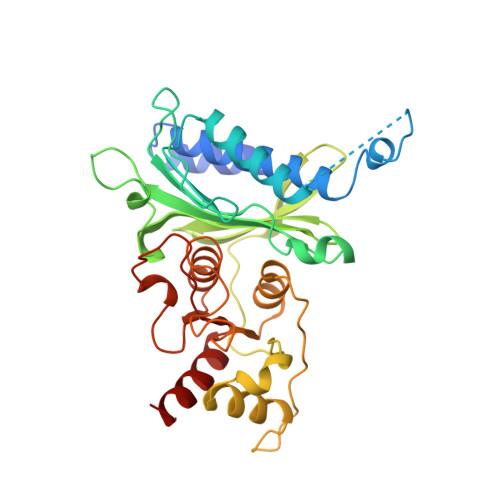
Structural aspects of the allosteric inhibition of fructose-1,6-bisphosphatase by AMP: the binding of both the substrate analogue 2,5-anhydro-D-glucitol 1,6-bisphosphate and catalytic metal ions monitored by X-ray crystallography.
Villeret, V., Huang, S., Zhang, Y., Lipscomb, W.N.(1995) Biochemistry 34: 4307-4315
- PubMed: 7703244
- DOI: https://doi.org/10.1021/bi00013a020
- Primary Citation of Related Structures:
1FPD, 1FPE, 1FPF, 1FPG - PubMed Abstract:
The crystal structures of the T form pig kidney fructose-1,6-bisphosphatase (EC 3.1.3.11) complexed with AMP, the substrate analogue 2,5-anhydro-D-glucitol 1,6-bisphosphate (AhG-1,6-P2), and Mn2+ at concentrations of 5, 15, 100, and 300 microM have been determined and refined at resolutions of 2.1-2.3 A to R factors which range from 0.180 to 0.195, respectively. Two metal ions per active site have been identified, one at a binding site of high affinity (metal site 1'), the second in a low affinity site (metal site 2'). The 1-phosphate group of the substrate analogue coordinates to the metal ion at site 1', but not at site 2'. In these four complexes, the distances between the two metal ions are all within 0.2 A of 4.3 A. In the previously determined R form structure of Fru-1,6-Pase complexed with AhG-1,6-P2 and Mn2+, there are also two metal ions in the active site at metal sites 1 and 2. The metal ion at site 1 is only 0.6 A displaced from the metal ion at site 1' in the T form and is also coordinated to the 1-phosphate group of AhG-1,6-P2. However, the second metal ion is located in two distinct sites which are 1.4 A apart in the T and R form structures. In the R form the Mn2+ at site 2 is coordinated to the 1-phosphate group of the substrate analogue. This metal ion is apparently required to orient the phosphate group for nucleophilic attack at the phosphorus center.(ABSTRACT TRUNCATED AT 250 WORDS)
Organizational Affiliation:
Gibbs Chemical Laboratory, Harvard University, Cambridge, Massachusetts 02138, USA.