Crystal structure of the neutral form of fructose 1,6-bisphosphatase complexed with regulatory inhibitor fructose 2,6-bisphosphate at 2.6-A resolution.
Liang, J.Y., Huang, S., Zhang, Y., Ke, H., Lipscomb, W.N.(1992) Proc Natl Acad Sci U S A 89: 2404-2408
- PubMed: 1312721
- DOI: https://doi.org/10.1073/pnas.89.6.2404
- Primary Citation of Related Structures:
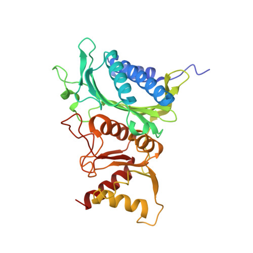
1FPB - PubMed Abstract:
The three-dimensional structure of the complex between fructose 1,6-bisphosphatase (EC 3.1.3.11) and the physiological inhibitor beta-D-fructose 2,6-bisphosphate (Fru-2,6-P2), an analogue of the substrate (fructose 1,6-bisphosphate), has been refined at 2.6-A resolution to a residual error (R) factor of 0.171. The rms deviations are 0.012 A and 2.88 degrees from ideal geometries of bond lengths and angles, respectively. The Fru-2,6-P2 occupies the active sites of both polypeptides C1 and C2 in the crystallographic asymmetric unit in the space group P3(2)21. The furanose and 6-phosphate of Fru-2,6-P2 are located at the fructose 6-phosphate site established earlier, and the 2-phosphate binds to the OH of Ser-124, the NH3+ of Lys-274, and the backbone NH of Gly-122 and Ser-123. Backbone displacements of 1 A occur for residues from Asp-121 to Asn-125. Model building of substrate alpha-D-Fru-1,6-P2 based on the binding structure of Fru-2,6-P2 in the active site shows interactions of the 1-phosphate with the backbone NH of Ser-123 and Ser-124. In the AMP sites, density peaks attributed to Fru-2,6-P2 are seen in C1 (and C4) but not in C2 (and C3). This minor binding of Fru-2,6-P2 to AMP sites partially explains the synergistic interaction between AMP and Fru-2,6-P2 and the protection of the AMP site from acetylation in the presence of Fru-2,6-P2. In the synergistic interaction between AMP and Fru-2,6-P2, inhibition of catalytic metal binding by the presence of Fru-2,6-P2 at the active site, and propagation of structural changes over some 28 A along beta-strand B3 from residues 121 to 125 in the active site to Lys-112 and Tyr-113 in the AMP site, as well as movement of helices across the interdimeric interfaces, may affect AMP binding and the subsequent R-to-T transition. In addition, occupancy of Fru-2,6-P2 at the AMP sites of C1 and C4 may favor binding of AMP to the remaining unoccupied AMP sites and thus promote the accompanying quaternary conformational changes.
Organizational Affiliation:
Gibbs Chemical Laboratory, Harvard University, Cambridge, MA 02138.