Insights into nucleotide signal transduction in nitrogenase: structure of an iron protein with MgADP bound.
Jang, S.B., Seefeldt, L.C., Peters, J.W.(2000) Biochemistry 39: 14745-14752
- PubMed: 11101289
- DOI: https://doi.org/10.1021/bi001705g
- Primary Citation of Related Structures:
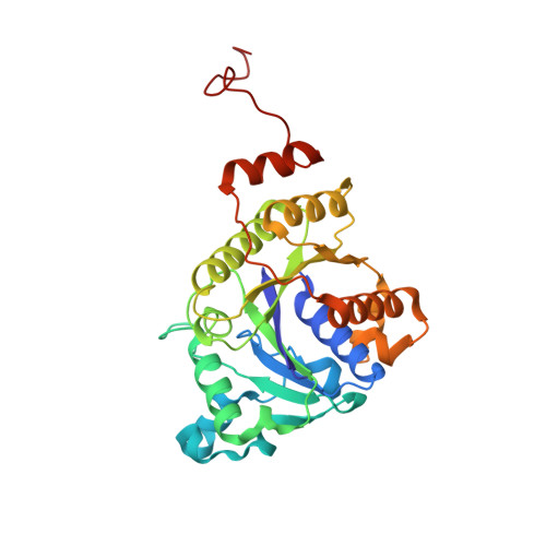
1FP6 - PubMed Abstract:
Coupling the energy of nucleoside triphosphate binding and hydrolysis to conformational changes is a common mechanism for a number of proteins with disparate cellular functions, including those involved in DNA replication, protein synthesis, and cell differentiation. Unique to this class of proteins is the dimeric Fe protein component of nitrogenase in which the binding and hydrolysis of MgATP controls intermolecular electron transfer and reduction of nitrogen to ammonia. In the work presented here, the MgADP-bound (or "off") conformational state of the nitrogenase Fe protein has been captured and a 2.15 A resolution X-ray crystal structure is presented. The structure described herein reveals likely mechanisms for long-range communication from the nucleotide-binding sites for controlling the affinity of association with the MoFe protein component. Two pathways, termed switches I and II, appear to be integral to this nucleotide signal transduction mechanism. In addition, the structure provides the basis for the changes in the biophysical properties of the [4Fe-4S] cluster observed when Fe protein binds nucleotides. The structure of the MgADP-bound Fe protein provides important insights into the respective contributions of nucleotide interaction and complex formation in defining the conformational states that are the keys to nitrogenase catalysis.
Organizational Affiliation:
Department of Chemistry and Biochemistry, Utah State University, Logan, Utah 84322, USA.