1FF2

CRYSTAL STRUCTURE OF THE C42D MUTANT OF AZOTOBACTER VINELANDII 7FE FERREDOXIN (FDI)


Experimental Data Snapshot

  • Method: X-RAY DIFFRACTION
  • Resolution: 2.30 Å
  • R-Value Free: 0.269 
  • R-Value Work: 0.217 
  • R-Value Observed: 0.217 

wwPDB Validation   3D Report Full Report


This is version 1.5 of the entry. See complete history


Literature

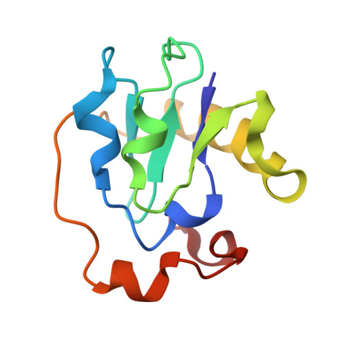
Structure of C42D Azotobacter vinelandii FdI. A Cys-X-X-Asp-X-X-Cys motif ligates an air-stable [4Fe-4S]2+/+ cluster

Jung, Y.S.Bonagura, C.A.Tilley, G.J.Gao-Sheridan, H.S.Armstrong, F.A.Stout, C.D.Burgess, B.K.

(2000) J Biol Chem 275: 36974-36983

  • DOI: https://doi.org/10.1074/jbc.M004947200
  • Primary Citation of Related Structures:  
    1FF2

  • PubMed Abstract: 

    All naturally occurring ferredoxins that have Cys-X-X-Asp-X-X-Cys motifs contain [4Fe-4S](2+/+) clusters that can be easily and reversibly converted to [3Fe-4S](+/0) clusters. In contrast, ferredoxins with unmodified Cys-X-X-Cys-X-X-Cys motifs assemble [4Fe-4S](2+/+) clusters that cannot be easily interconverted with [3Fe-4S](+/0) clusters. In this study we changed the central cysteine of the Cys(39)-X-X-Cys(42)-X-X-Cys(45) of Azotobacter vinelandii FdI, which coordinates its [4Fe-4S](2+/+) cluster, into an aspartate. UV-visible, EPR, and CD spectroscopies, metal analysis, and x-ray crystallography show that, like native FdI, aerobically purified C42D FdI is a seven-iron protein retaining its [4Fe-4S](2+/+) cluster with monodentate aspartate ligation to one iron. Unlike known clusters of this type the reduced [4Fe-4S](+) cluster of C42D FdI exhibits only an S = 1/2 EPR with no higher spin signals detected. The cluster shows only a minor change in reduction potential relative to the native protein. All attempts to convert the cluster to a 3Fe cluster using conventional methods of oxygen or ferricyanide oxidation or thiol exchange were not successful. The cluster conversion was ultimately accomplished using a new electrochemical method. Hydrophobic and electrostatic interaction and the lack of Gly residues adjacent to the Asp ligand explain the remarkable stability of this cluster.


  • Organizational Affiliation

    Department of Molecular Biology and Biochemistry, University of California, Irvine, California 92697, USA.


Macromolecules
Find similar proteins by:  (by identity cutoff)  |  3D Structure
Entity ID: 1
MoleculeChains Sequence LengthOrganismDetailsImage
FERREDOXIN I106Azotobacter vinelandiiMutation(s): 1 
UniProt
Find proteins for P00214 (Azotobacter vinelandii)
Explore P00214 
Go to UniProtKB:  P00214
Entity Groups  
Sequence Clusters30% Identity50% Identity70% Identity90% Identity95% Identity100% Identity
UniProt GroupP00214
Sequence Annotations
Expand
  • Reference Sequence
Small Molecules
Ligands 2 Unique
IDChains Name / Formula / InChI Key2D Diagram3D Interactions
SF4
Query on SF4

Download Ideal Coordinates CCD File 
B [auth A]IRON/SULFUR CLUSTER
Fe4 S4
LJBDFODJNLIPKO-UHFFFAOYSA-N
F3S
Query on F3S

Download Ideal Coordinates CCD File 
C [auth A]FE3-S4 CLUSTER
Fe3 S4
FCXHZBQOKRZXKS-UHFFFAOYSA-N
Experimental Data & Validation

Experimental Data

  • Method: X-RAY DIFFRACTION
  • Resolution: 2.30 Å
  • R-Value Free: 0.269 
  • R-Value Work: 0.217 
  • R-Value Observed: 0.217 
  • Space Group: P 41 21 2
Unit Cell:
Length ( Å )Angle ( ˚ )
a = 55.3α = 90
b = 55.3β = 90
c = 90.59γ = 90
Software Package:
Software NamePurpose
DENZOdata reduction
X-PLORmodel building
X-PLORrefinement
SCALEPACKdata scaling
X-PLORphasing

Structure Validation

View Full Validation Report


Entry History 

Deposition Data

Revision History  (Full details and data files)

  • Version 1.0: 2000-08-21
    Type: Initial release
  • Version 1.1: 2008-04-27
    Changes: Version format compliance
  • Version 1.2: 2011-07-13
    Changes: Version format compliance
  • Version 1.3: 2017-10-04
    Changes: Refinement description
  • Version 1.4: 2021-11-03
    Changes: Database references, Derived calculations
  • Version 1.5: 2024-02-07
    Changes: Data collection