1FA0

STRUCTURE OF YEAST POLY(A) POLYMERASE BOUND TO MANGANATE AND 3'-DATP


Experimental Data Snapshot

  • Method: X-RAY DIFFRACTION
  • Resolution: 2.60 Å
  • R-Value Free: 0.278 
  • R-Value Work: 0.233 
  • R-Value Observed: 0.233 

wwPDB Validation   3D Report Full Report


This is version 1.3 of the entry. See complete history


Literature

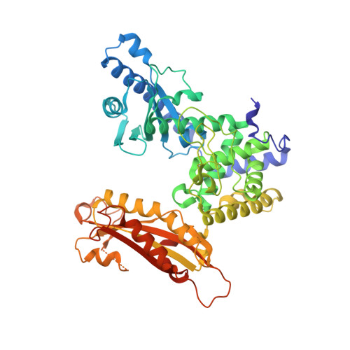
Structure of yeast poly(A) polymerase alone and in complex with 3'-dATP.

Bard, J.Zhelkovsky, A.M.Helmling, S.Earnest, T.N.Moore, C.L.Bohm, A.

(2000) Science 289: 1346-1349

  • DOI: https://doi.org/10.1126/science.289.5483.1346
  • Primary Citation of Related Structures:  
    1FA0

  • PubMed Abstract: 

    Polyadenylate [poly(A)] polymerase (PAP) catalyzes the addition of a polyadenosine tail to almost all eukaryotic messenger RNAs (mRNAs). The crystal structure of the PAP from Saccharomyces cerevisiae (Pap1) has been solved to 2.6 angstroms, both alone and in complex with 3'-deoxyadenosine triphosphate (3'-dATP). Like other nucleic acid polymerases, Pap1 is composed of three domains that encircle the active site. The arrangement of these domains, however, is quite different from that seen in polymerases that use a template to select and position their incoming nucleotides. The first two domains are functionally analogous to polymerase palm and fingers domains. The third domain is attached to the fingers domain and is known to interact with the single-stranded RNA primer. In the nucleotide complex, two molecules of 3'-dATP are bound to Pap1. One occupies the position of the incoming base, prior to its addition to the mRNA chain. The other is believed to occupy the position of the 3' end of the mRNA primer.


  • Organizational Affiliation

    Boston Biomedical Research Institute, 64 Grove Street, Watertown, MA 02472, USA.


Macromolecules
Find similar proteins by:  (by identity cutoff)  |  3D Structure
Entity ID: 1
MoleculeChains Sequence LengthOrganismDetailsImage
POLY(A)-POLYMERASE
A, B
537Saccharomyces cerevisiaeMutation(s): 0 
EC: 2.7.7.19
UniProt
Find proteins for P29468 (Saccharomyces cerevisiae (strain ATCC 204508 / S288c))
Explore P29468 
Go to UniProtKB:  P29468
Entity Groups  
Sequence Clusters30% Identity50% Identity70% Identity90% Identity95% Identity100% Identity
UniProt GroupP29468
Sequence Annotations
Expand
  • Reference Sequence
Small Molecules
Ligands 4 Unique
IDChains Name / Formula / InChI Key2D Diagram3D Interactions
3AT
Query on 3AT

Download Ideal Coordinates CCD File 
E [auth A],
J [auth B]
3'-DEOXYADENOSINE-5'-TRIPHOSPHATE
C10 H16 N5 O12 P3
NLIHPCYXRYQPSD-BAJZRUMYSA-N
3AD
Query on 3AD

Download Ideal Coordinates CCD File 
F [auth A],
K [auth B]
3'-DEOXYADENOSINE
C10 H13 N5 O3
OFEZSBMBBKLLBJ-BAJZRUMYSA-N
POP
Query on POP

Download Ideal Coordinates CCD File 
G [auth A]PYROPHOSPHATE 2-
H2 O7 P2
XPPKVPWEQAFLFU-UHFFFAOYSA-L
MN
Query on MN

Download Ideal Coordinates CCD File 
C [auth A],
D [auth A],
H [auth B],
I [auth B]
MANGANESE (II) ION
Mn
WAEMQWOKJMHJLA-UHFFFAOYSA-N
Experimental Data & Validation

Experimental Data

  • Method: X-RAY DIFFRACTION
  • Resolution: 2.60 Å
  • R-Value Free: 0.278 
  • R-Value Work: 0.233 
  • R-Value Observed: 0.233 
  • Space Group: P 21 21 21
Unit Cell:
Length ( Å )Angle ( ˚ )
a = 73.8α = 90
b = 109.1β = 90
c = 238.5γ = 90
Software Package:
Software NamePurpose
CNSrefinement
DENZOdata reduction
SCALEPACKdata scaling
CNSphasing

Structure Validation

View Full Validation Report


Entry History 

Deposition Data

Revision History  (Full details and data files)

  • Version 1.0: 2000-08-30
    Type: Initial release
  • Version 1.1: 2008-04-27
    Changes: Version format compliance
  • Version 1.2: 2011-07-13
    Changes: Version format compliance
  • Version 1.3: 2024-02-07
    Changes: Data collection, Database references, Derived calculations