1F57

CARBOXYPEPTIDASE A COMPLEX WITH D-CYSTEINE AT 1.75 A


Experimental Data Snapshot

  • Method: X-RAY DIFFRACTION
  • Resolution: 1.75 Å
  • R-Value Free: 0.164 
  • R-Value Work: 0.137 
  • R-Value Observed: 0.137 

wwPDB Validation   3D Report Full Report


This is version 1.4 of the entry. See complete history


Literature

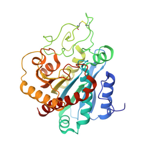
Crystal structure of carboxypeptidase A complexed with D-cysteine at 1.75 A - inhibitor-induced conformational changes.

van Aalten, D.M.Chong, C.R.Joshua-Tor, L.

(2000) Biochemistry 39: 10082-10089

  • DOI: https://doi.org/10.1021/bi000952h
  • Primary Citation of Related Structures:  
    1F57

  • PubMed Abstract: 

    D-Cysteine differs from the antiarthritis drug D-penicillamine by only two methyl groups on the beta-carbon yet inhibits carboxypeptidase A (CPD) by a distinct mechanism: D-cysteine binds tightly to the active site zinc, while D-penicillamine catalyzes metal removal. To investigate the structural basis for this difference, we solved the crystal structure of carboxypeptidase A complexed with D-cysteine (D-Cys) at 1.75-A resolution. D-Cys binds the active site zinc with a sulfur ligand and forms additional interactions with surrounding side chains of the enzyme. The structure explains the difference in potency between D-Cys and L-Cys and provides insight into the mechanism of D-penicillamine inhibition. D-Cys binding induces a concerted motion of the side chains around the zinc ion, similar to that found in other carboxypeptidase-inhibitor crystal structures and along a limited path. Analysis of concerted motions of CPD and CPD-inhibitor crystal structures reveals a clustering of these structures into distinct groups. Using the restricted conformational flexibility of a drug target in this type of analysis could greatly enhance efficiency in drug design.


  • Organizational Affiliation

    W. M. Keck Structural Biology, Cold Spring Harbor Laboratory, Cold Spring Harbor, NY 11724, USA.


Macromolecules
Find similar proteins by:  (by identity cutoff)  |  3D Structure
Entity ID: 1
MoleculeChains Sequence LengthOrganismDetailsImage
CARBOXYPEPTIDASE A307Bos taurusMutation(s): 0 
EC: 3.4.17.1
UniProt
Find proteins for P00730 (Bos taurus)
Explore P00730 
Go to UniProtKB:  P00730
Entity Groups  
Sequence Clusters30% Identity50% Identity70% Identity90% Identity95% Identity100% Identity
UniProt GroupP00730
Sequence Annotations
Expand
  • Reference Sequence
Small Molecules
Ligands 2 Unique
IDChains Name / Formula / InChI Key2D Diagram3D Interactions
DCY
Query on DCY

Download Ideal Coordinates CCD File 
C [auth A]D-CYSTEINE
C3 H7 N O2 S
XUJNEKJLAYXESH-UWTATZPHSA-N
ZN
Query on ZN

Download Ideal Coordinates CCD File 
B [auth A]ZINC ION
Zn
PTFCDOFLOPIGGS-UHFFFAOYSA-N
Binding Affinity Annotations 
IDSourceBinding Affinity
DCY PDBBind:  1F57 Ki: 2300 (nM) from 1 assay(s)
BindingDB:  1F57 Ki: 2300 (nM) from 1 assay(s)
Binding MOAD:  1F57 Ki: 2300 (nM) from 1 assay(s)
Experimental Data & Validation

Experimental Data

  • Method: X-RAY DIFFRACTION
  • Resolution: 1.75 Å
  • R-Value Free: 0.164 
  • R-Value Work: 0.137 
  • R-Value Observed: 0.137 
  • Space Group: P 1 21 1
Unit Cell:
Length ( Å )Angle ( ˚ )
a = 44.389α = 90
b = 57.712β = 101.62
c = 58.193γ = 90
Software Package:
Software NamePurpose
DENZOdata reduction
SCALEPACKdata scaling
AMoREphasing
CNSrefinement

Structure Validation

View Full Validation Report


Entry History 

Deposition Data

Revision History  (Full details and data files)

  • Version 1.0: 2000-09-06
    Type: Initial release
  • Version 1.1: 2008-04-27
    Changes: Version format compliance
  • Version 1.2: 2011-07-13
    Changes: Version format compliance
  • Version 1.3: 2018-01-31
    Changes: Advisory, Experimental preparation
  • Version 1.4: 2023-08-09
    Changes: Advisory, Data collection, Database references, Derived calculations, Refinement description