The three-dimensional structure of the Nudix enzyme diadenosine tetraphosphate hydrolase from Lupinus angustifolius L.
Swarbrick, J.D., Bashtannyk, T., Maksel, D., Zhang, X.R., Blackburn, G.M., Gayler, K.R., Gooley, P.R.(2000) J Mol Biol 302: 1165-1177
- PubMed: 11183782
- DOI: https://doi.org/10.1006/jmbi.2000.4085
- Primary Citation of Related Structures:
1F3Y - PubMed Abstract:
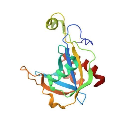
The solution structure of diadenosine 5',5'''-P1,P4-tetraphosphate hydrolase from Lupinus angustifolius L., an enzyme of the Nudix family, has been determined by heteronuclear NMR, using a torsion angle dynamics/simulated annealing protocol based on approximately 12 interresidue NOEs per residue. The structure represents the first Ap4A hydrolase to be determined, and sequence homology suggests that other members will have the same fold. The family of structures shows a well-defined fold comprised of a central four-stranded mixed beta-sheet, a two-stranded antiparallel beta-sheet and three helices (alphaI, alphaIII, alphaIV). The root-mean-squared deviation for the backbone (C',O,N,Calpha) of the rigid parts (residues 9 to 75, 97 to 115, 125 to 160) of the protein is 0.32 A. Several regions, however, show lower definition, particularly an isolated helix (alphaII) that connects two strands of the central sheet. This poor definition is mainly due to a lack of long-range NOEs between alphaII and other parts of the protein. Mapping conserved residues outside of the Nudix signature and those sensitive to an Ap4A analogue suggests that the adenosine-ribose moiety of the substrate binds into a large cleft above the four-stranded beta-sheet. Four conserved glutamate residues (Glu55, Glu58, Glu59 and Glu125) form a cluster that most likely ligates an essential magnesium ion, however, Gly41 also an expected magnesium ligand, is distant from this cluster.
Organizational Affiliation:
Department of Biochemistry and Molecular Biology, University of Melbourne, Parkville, Victoria, Australia.