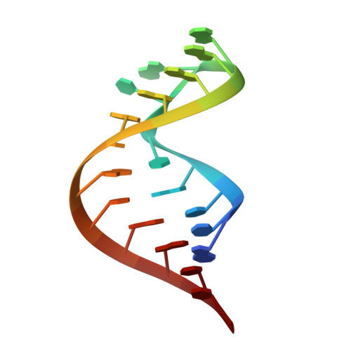
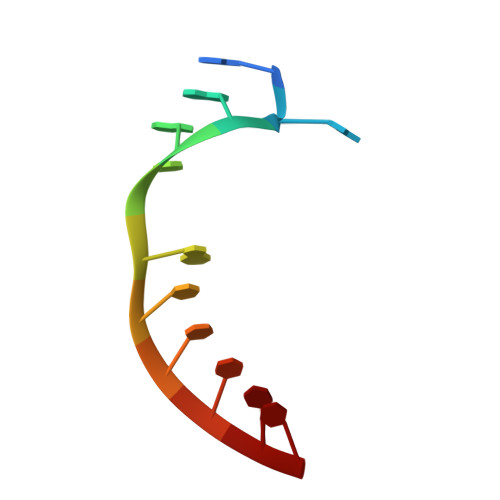
The 1.3 A crystal structure of a biotin-binding pseudoknot and the basis for RNA molecular recognition.
Nix, J., Sussman, D., Wilson, C.(2000) J Mol Biol 296: 1235-1244
- PubMed: 10698630
- DOI: https://doi.org/10.1006/jmbi.2000.3539
- Primary Citation of Related Structures:
1F27 - PubMed Abstract:
A pseudoknot-containing aptamer isolated from a pool of random sequence molecules has been shown previously to represent an optimal RNA solution to the problem of binding biotin. The affinity of this RNA molecule is nonetheless orders of magnitude weaker than that of its highly evolved protein analogs, avidin and streptavidin. To understand the structural basis for biotin binding and to compare directly strategies for ligand recognition available to proteins and RNA molecules, we have determined the 1.3 A crystal structure of the aptamer complexed with its ligand. Biotin is bound at the interface between the pseudoknot's stacked helices in a pocket defined almost entirely by base-paired nucleotides. In comparison to the protein avidin, the aptamer packs more tightly around the biotin headgroup and makes fewer contacts with its fatty acid tail. Whereas biotin is deeply buried within the hydrophobic core in the avidin complex, the aptamer relies on a combination of hydrated magnesium ions and immobilized water molecules to surround its ligand. In addition to demonstrating fundamentally different approaches to molecular recognition by proteins and RNA, the structure provides general insight into the mechanisms by which RNA function is mediated by divalent metals.
Organizational Affiliation:
Department of Biology and Center for the Molecular Biology of RNA, Sinsheimer Laboratories, University of California at Santa Cruz, Santa Cruz, CA 95064, USA. nix@biology.ucsc.edu