1F0K

THE 1.9 ANGSTROM CRYSTAL STRUCTURE OF E. COLI MURG


Experimental Data Snapshot

  • Method: X-RAY DIFFRACTION
  • Resolution: 1.90 Å
  • R-Value Free: 0.247 
  • R-Value Work: 0.220 

wwPDB Validation   3D Report Full Report


This is version 1.3 of the entry. See complete history


Literature

The 1.9 A crystal structure of Escherichia coli MurG, a membrane-associated glycosyltransferase involved in peptidoglycan biosynthesis.

Ha, S.Walker, D.Shi, Y.Walker, S.

(2000) Protein Sci 9: 1045-1052

  • DOI: https://doi.org/10.1110/ps.9.6.1045
  • Primary Citation of Related Structures:  
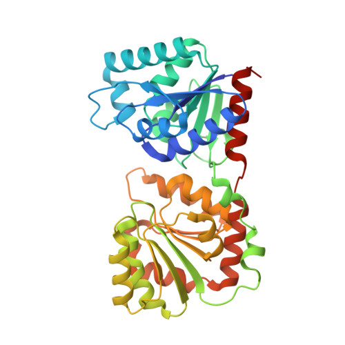
    1F0K

  • PubMed Abstract: 

    The 1.9 A X-ray structure of a membrane-associated glycosyltransferase involved in peptidoglycan biosynthesis is reported. This enzyme, MurG, contains two alpha/beta open sheet domains separated by a deep cleft. Structural analysis suggests that the C-terminal domain contains the UDP-GlcNAc binding site while the N-terminal domain contains the acceptor binding site and likely membrane association site. Combined with sequence data from other MurG homologs, this structure provides insight into the residues that are important in substrate binding and catalysis. We have also noted that a conserved region found in many UDP-sugar transferases maps to a beta/alpha/beta/alpha supersecondary structural motif in the donor binding region of MurG, an observation that may be helpful in glycosyltransferase structure prediction. The identification of a conserved structural motif involved in donor binding in different UDP-sugar transferases also suggests that it may be possible to identify--and perhaps alter--the residues that help determine donor specificity.


  • Organizational Affiliation

    Department of Chemistry, Princeton University, New Jersey 08544, USA.


Macromolecules
Find similar proteins by:  (by identity cutoff)  |  3D Structure
Entity ID: 1
MoleculeChains Sequence LengthOrganismDetailsImage
UDP-N-ACETYLGLUCOSAMINE-N-ACETYLMURAMYL-(PENTAPEPTIDE) PYROPHOSPHORYL-UNDECAPRENOL N-ACETYLGLUCOSAMINE TRANSFERASE
A, B
364Escherichia coliMutation(s): 0 
EC: 2.4.1
Membrane Entity: Yes 
UniProt
Find proteins for P17443 (Escherichia coli (strain K12))
Explore P17443 
Go to UniProtKB:  P17443
Entity Groups  
Sequence Clusters30% Identity50% Identity70% Identity90% Identity95% Identity100% Identity
UniProt GroupP17443
Sequence Annotations
Expand
  • Reference Sequence
Small Molecules
Ligands 1 Unique
IDChains Name / Formula / InChI Key2D Diagram3D Interactions
SO4
Query on SO4

Download Ideal Coordinates CCD File 
C [auth B]SULFATE ION
O4 S
QAOWNCQODCNURD-UHFFFAOYSA-L
Experimental Data & Validation

Experimental Data

  • Method: X-RAY DIFFRACTION
  • Resolution: 1.90 Å
  • R-Value Free: 0.247 
  • R-Value Work: 0.220 
  • Space Group: P 1
Unit Cell:
Length ( Å )Angle ( ˚ )
a = 60.613α = 64.29
b = 66.356β = 83.52
c = 67.902γ = 65.45
Software Package:
Software NamePurpose
MLPHAREphasing
CNSrefinement
DENZOdata reduction
SCALEPACKdata scaling

Structure Validation

View Full Validation Report


Entry History 

Deposition Data

Revision History  (Full details and data files)

  • Version 1.0: 2000-07-27
    Type: Initial release
  • Version 1.1: 2008-04-27
    Changes: Version format compliance
  • Version 1.2: 2011-07-13
    Changes: Version format compliance
  • Version 1.3: 2024-02-07
    Changes: Data collection, Database references, Derived calculations