1F05

CRYSTAL STRUCTURE OF HUMAN TRANSALDOLASE


Experimental Data Snapshot

  • Method: X-RAY DIFFRACTION
  • Resolution: 2.45 Å
  • R-Value Free: 0.258 
  • R-Value Work: 0.225 
  • R-Value Observed: 0.225 

wwPDB Validation   3D Report Full Report


This is version 1.5 of the entry. See complete history


Literature

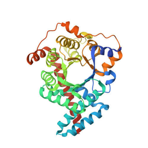
The three-dimensional structure of human transaldolase.

Thorell, S.Gergely Jr., P.Banki, K.Perl, A.Schneider, G.

(2000) FEBS Lett 475: 205-208

  • DOI: https://doi.org/10.1016/s0014-5793(00)01658-6
  • Primary Citation of Related Structures:  
    1F05

  • PubMed Abstract: 

    The crystal structure of human transaldolase has been determined to 2.45 A resolution. The enzyme folds into an alpha/beta barrel structure and is thus similar in structure to other class I aldolases. Structure-based sequence alignment of available sequences of the transaldolase subfamily reveals that eight active site residues are invariant in the whole subfamily. Other invariant residues are mainly involved in the formation of the hydrophobic core of the enzyme. Noteworthy is a hydrophobic cluster consisting of five invariant residues. Human transaldolase has been implicated as an autoantigen in multiple sclerosis and four immunodominant peptide segments are located at the surface of the enzyme, accessible to autoantibodies.


  • Organizational Affiliation

    Department of Medical Biochemistry and Biophysics, Karolinska Institutet, Stockholm, Sweden.


Macromolecules
Find similar proteins by:  (by identity cutoff)  |  3D Structure
Entity ID: 1
MoleculeChains Sequence LengthOrganismDetailsImage
TRANSALDOLASE
A, B
337Homo sapiensMutation(s): 0 
EC: 2.2.1.2
UniProt & NIH Common Fund Data Resources
Find proteins for P37837 (Homo sapiens)
Explore P37837 
Go to UniProtKB:  P37837
PHAROS:  P37837
GTEx:  ENSG00000177156 
Entity Groups  
Sequence Clusters30% Identity50% Identity70% Identity90% Identity95% Identity100% Identity
UniProt GroupP37837
Sequence Annotations
Expand
  • Reference Sequence
Experimental Data & Validation

Experimental Data

  • Method: X-RAY DIFFRACTION
  • Resolution: 2.45 Å
  • R-Value Free: 0.258 
  • R-Value Work: 0.225 
  • R-Value Observed: 0.225 
  • Space Group: P 1 21 1
Unit Cell:
Length ( Å )Angle ( ˚ )
a = 45.57α = 90
b = 113.57β = 101.37
c = 69.19γ = 90
Software Package:
Software NamePurpose
AMoREphasing
CNSrefinement
MAR345data collection
CCP4data scaling

Structure Validation

View Full Validation Report


Entry History 

Deposition Data

Revision History  (Full details and data files)

  • Version 1.0: 2000-07-13
    Type: Initial release
  • Version 1.1: 2008-04-27
    Changes: Version format compliance
  • Version 1.2: 2011-07-13
    Changes: Version format compliance
  • Version 1.3: 2017-10-04
    Changes: Data collection, Refinement description
  • Version 1.4: 2018-01-31
    Changes: Experimental preparation
  • Version 1.5: 2023-08-09
    Changes: Data collection, Database references, Refinement description