1EZX

CRYSTAL STRUCTURE OF A SERPIN:PROTEASE COMPLEX


Experimental Data Snapshot

  • Method: X-RAY DIFFRACTION
  • Resolution: 2.60 Å
  • R-Value Free: 0.239 
  • R-Value Work: 0.205 

wwPDB Validation   3D Report Full Report


This is version 1.2 of the entry. See complete history


Literature

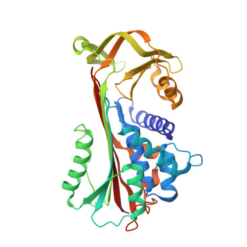
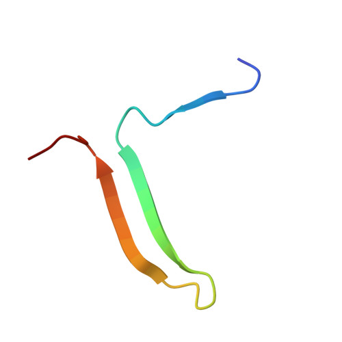
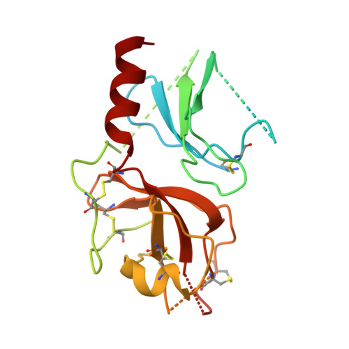
Structure of a serpin-protease complex shows inhibition by deformation.

Huntington, J.A.Read, R.J.Carrell, R.W.

(2000) Nature 407: 923-926

  • DOI: https://doi.org/10.1038/35038119
  • Primary Citation of Related Structures:  
    1EZX

  • PubMed Abstract: 

    The serpins have evolved to be the predominant family of serine-protease inhibitors in man. Their unique mechanism of inhibition involves a profound change in conformation, although the nature and significance of this change has been controversial. Here we report the crystallographic structure of a typical serpin-protease complex and show the mechanism of inhibition. The conformational change is initiated by reaction of the active serine of the protease with the reactive centre of the serpin. This cleaves the reactive centre, which then moves 71 A to the opposite pole of the serpin, taking the tethered protease with it. The tight linkage of the two molecules and resulting overlap of their structures does not affect the hyperstable serpin, but causes a surprising 37% loss of structure in the protease. This is induced by the plucking of the serine from its active site, together with breakage of interactions formed during zymogen activation. The disruption of the catalytic site prevents the release of the protease from the complex, and the structural disorder allows its proteolytic destruction. It is this ability of the conformational mechanism to crush as well as inhibit proteases that provides the serpins with their selective advantage.


  • Organizational Affiliation

    Department of Haematology, University of Cambridge, Wellcome Trust Centre for Molecular Mechanisms in Disease, Cambridge Institute for Medical Research, UK. rwc1000@cam.ac.uk


Macromolecules
Find similar proteins by:  (by identity cutoff)  |  3D Structure
Entity ID: 1
MoleculeChains Sequence LengthOrganismDetailsImage
ALPHA-1-ANTITRYPSIN335Homo sapiensMutation(s): 0 
UniProt & NIH Common Fund Data Resources
Find proteins for P01009 (Homo sapiens)
Explore P01009 
Go to UniProtKB:  P01009
PHAROS:  P01009
GTEx:  ENSG00000197249 
Entity Groups  
Sequence Clusters30% Identity50% Identity70% Identity90% Identity95% Identity100% Identity
UniProt GroupP01009
Sequence Annotations
Expand
  • Reference Sequence
Find similar proteins by:  (by identity cutoff)  |  3D Structure
Entity ID: 2
MoleculeChains Sequence LengthOrganismDetailsImage
ALPHA-1-ANTITRYPSIN36Homo sapiensMutation(s): 0 
UniProt & NIH Common Fund Data Resources
Find proteins for P01009 (Homo sapiens)
Explore P01009 
Go to UniProtKB:  P01009
PHAROS:  P01009
GTEx:  ENSG00000197249 
Entity Groups  
Sequence Clusters30% Identity50% Identity70% Identity90% Identity95% Identity100% Identity
UniProt GroupP01009
Sequence Annotations
Expand
  • Reference Sequence
Find similar proteins by:  (by identity cutoff)  |  3D Structure
Entity ID: 3
MoleculeChains Sequence LengthOrganismDetailsImage
TRYPSIN243Bos taurusMutation(s): 0 
EC: 3.4.21.4
UniProt
Find proteins for P00760 (Bos taurus)
Explore P00760 
Go to UniProtKB:  P00760
Entity Groups  
Sequence Clusters30% Identity50% Identity70% Identity90% Identity95% Identity100% Identity
UniProt GroupP00760
Sequence Annotations
Expand
  • Reference Sequence
Experimental Data & Validation

Experimental Data

  • Method: X-RAY DIFFRACTION
  • Resolution: 2.60 Å
  • R-Value Free: 0.239 
  • R-Value Work: 0.205 
  • Space Group: C 2 2 21
Unit Cell:
Length ( Å )Angle ( ˚ )
a = 63.234α = 90
b = 171.295β = 90
c = 145.78γ = 90
Software Package:
Software NamePurpose
MOSFLMdata reduction
SCALAdata scaling
AMoREphasing
CNSrefinement
CCP4data scaling

Structure Validation

View Full Validation Report


Entry History 

Deposition Data

Revision History  (Full details and data files)

  • Version 1.0: 2000-10-25
    Type: Initial release
  • Version 1.1: 2008-04-27
    Changes: Version format compliance
  • Version 1.2: 2011-07-13
    Changes: Version format compliance