1EUS

SIALIDASE COMPLEXED WITH 2-DEOXY-2,3-DEHYDRO-N-ACETYLNEURAMINIC ACID


Experimental Data Snapshot

  • Method: X-RAY DIFFRACTION
  • Resolution: 2.00 Å
  • R-Value Work: 0.159 
  • R-Value Observed: 0.159 

wwPDB Validation   3D Report Full Report


This is version 1.4 of the entry. See complete history


Literature

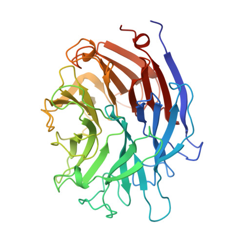
The three domains of a bacterial sialidase: a beta-propeller, an immunoglobulin module and a galactose-binding jelly-roll.

Gaskell, A.Crennell, S.Taylor, G.

(1995) Structure 3: 1197-1205

  • DOI: https://doi.org/10.1016/s0969-2126(01)00255-6
  • Primary Citation of Related Structures:  
    1EUR, 1EUS, 1EUT

  • PubMed Abstract: 

    Sialidases, or neuraminidases, have been implicated in the pathogenesis of many diseases, but are also produced by many non-pathogenic bacteria. Bacterial sialidases are very variable in size, often possessing domains in addition to the catalytic domain. The sialidase from the non-pathogenic soil bacterium Micromonospora viridifaciens is secreted in two forms with molecular weights of 41 kDa or 68 kDa, depending on the nature of the carbohydrate used to induce expression.


  • Organizational Affiliation

    School of Biology and Biochemistry, University of Bath, UK.


Macromolecules
Find similar proteins by:  (by identity cutoff)  |  3D Structure
Entity ID: 1
MoleculeChains Sequence LengthOrganismDetailsImage
SIALIDASE365Micromonospora viridifaciensMutation(s): 0 
EC: 3.2.1.18
UniProt
Find proteins for Q02834 (Micromonospora viridifaciens)
Explore Q02834 
Go to UniProtKB:  Q02834
Entity Groups  
Sequence Clusters30% Identity50% Identity70% Identity90% Identity95% Identity100% Identity
UniProt GroupQ02834
Sequence Annotations
Expand
  • Reference Sequence
Small Molecules
Ligands 1 Unique
IDChains Name / Formula / InChI Key2D Diagram3D Interactions
DAN
Query on DAN

Download Ideal Coordinates CCD File 
B [auth A]2-DEOXY-2,3-DEHYDRO-N-ACETYL-NEURAMINIC ACID
C11 H17 N O8
JINJZWSZQKHCIP-UFGQHTETSA-N
Experimental Data & Validation

Experimental Data

  • Method: X-RAY DIFFRACTION
  • Resolution: 2.00 Å
  • R-Value Work: 0.159 
  • R-Value Observed: 0.159 
  • Space Group: P 21 21 21
Unit Cell:
Length ( Å )Angle ( ˚ )
a = 48α = 90
b = 82.86β = 90
c = 84.94γ = 90
Software Package:
Software NamePurpose
X-PLORmodel building
X-PLORrefinement
XDSdata reduction
X-PLORphasing

Structure Validation

View Full Validation Report


Entry History 

Deposition Data

Revision History  (Full details and data files)

  • Version 1.0: 1997-01-11
    Type: Initial release
  • Version 1.1: 2008-03-24
    Changes: Version format compliance
  • Version 1.2: 2011-07-13
    Changes: Version format compliance
  • Version 1.3: 2020-07-29
    Type: Remediation
    Reason: Carbohydrate remediation
    Changes: Data collection, Derived calculations, Other
  • Version 1.4: 2024-02-07
    Changes: Data collection, Database references, Structure summary