1EPP

ENDOTHIA ASPARTIC PROTEINASE (ENDOTHIAPEPSIN) COMPLEXED WITH PD-130,693 (MAS PHE LYS+MTF STA MBA)


Experimental Data Snapshot

  • Method: X-RAY DIFFRACTION
  • Resolution: 1.90 Å
  • R-Value Observed: 0.160 

wwPDB Validation   3D Report Full Report


This is version 1.4 of the entry. See complete history


Literature

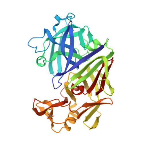
Analyses of ligand binding in five endothiapepsin crystal complexes and their use in the design and evaluation of novel renin inhibitors.

Lunney, E.A.Hamilton, H.W.Hodges, J.C.Kaltenbronn, J.S.Repine, J.T.Badasso, M.Cooper, J.B.Dealwis, C.Wallace, B.A.Lowther, W.T.

(1993) J Med Chem 36: 3809-3820

  • DOI: https://doi.org/10.1021/jm00076a008
  • Primary Citation of Related Structures:  
    1EPP, 1EPQ

  • PubMed Abstract: 

    Five renin inhibitors were cocrystallized with endothiapepsin, a fungal enzyme homologous to renin. Crystal structures of inhibitor-bound complexes have provided invaluable insight regarding the three-dimensional structure of the aspartic proteinase family of enzymes, as well as the steric and polar interactions that occur between the proteins and the bound ligands. Beyond this, subtleties of binding have been revealed, including multiple subsite binding modes and subsite interdependencies. This information has been applied in the design of novel potent renin inhibitors and in the understanding of structure-activity relationships and enzyme selectivities.


  • Organizational Affiliation

    Parke-Davis Pharmaceutical Research, Division of Warner-Lambert Company, Ann Arbor, Michigan 48105-2430.


Macromolecules
Find similar proteins by:  (by identity cutoff)  |  3D Structure
Entity ID: 1
MoleculeChains Sequence LengthOrganismDetailsImage
ENDOTHIAPEPSINA [auth E]330Cryphonectria parasiticaMutation(s): 0 
EC: 3.4.23.22
UniProt
Find proteins for P11838 (Cryphonectria parasitica)
Explore P11838 
Go to UniProtKB:  P11838
Entity Groups  
Sequence Clusters30% Identity50% Identity70% Identity90% Identity95% Identity100% Identity
UniProt GroupP11838
Sequence Annotations
Expand
  • Reference Sequence
Small Molecules
Ligands 2 Unique
IDChains Name / Formula / InChI Key2D Diagram3D Interactions
1Z1
Query on 1Z1

Download Ideal Coordinates CCD File 
D [auth E]N-(dimethylsulfamoyl)-L-phenylalanyl-N-[(1S,2S)-2-hydroxy-4-{[(2S)-2-methylbutyl]amino}-1-(2-methylpropyl)-4-oxobutyl]-N~6~-(methylcarbamothioyl)-L-lysinamide
C32 H57 N7 O6 S2
GBPRCFWBWHGSJH-BLVAWXTGSA-N
SO4
Query on SO4

Download Ideal Coordinates CCD File 
B [auth E],
C [auth E]
SULFATE ION
O4 S
QAOWNCQODCNURD-UHFFFAOYSA-L
Binding Affinity Annotations 
IDSourceBinding Affinity
1Z1 Binding MOAD:  1EPP Ki: 69 (nM) from 1 assay(s)
Experimental Data & Validation

Experimental Data

  • Method: X-RAY DIFFRACTION
  • Resolution: 1.90 Å
  • R-Value Observed: 0.160 
  • Space Group: P 1 21 1
Unit Cell:
Length ( Å )Angle ( ˚ )
a = 43.1α = 90
b = 75.6β = 97.2
c = 42.9γ = 90
Software Package:
Software NamePurpose
RESTRAINrefinement

Structure Validation

View Full Validation Report


Entry History 

Deposition Data

Revision History  (Full details and data files)

  • Version 1.0: 1994-12-20
    Type: Initial release
  • Version 1.1: 2008-03-10
    Changes: Version format compliance
  • Version 1.2: 2011-07-13
    Changes: Atomic model, Database references, Derived calculations, Non-polymer description, Structure summary, Version format compliance
  • Version 1.3: 2012-12-12
    Changes: Other
  • Version 1.4: 2017-11-29
    Changes: Derived calculations, Other