1EPB

STRUCTURE OF THE EPIDIDYMAL RETINOIC ACID-BINDING PROTEIN AT 2.1 ANGSTROMS RESOLUTION


Experimental Data Snapshot

  • Method: X-RAY DIFFRACTION
  • Resolution: 2.20 Å
  • R-Value Work: 0.182 
  • R-Value Observed: 0.182 

wwPDB Validation   3D Report Full Report


This is version 1.4 of the entry. See complete history


Literature

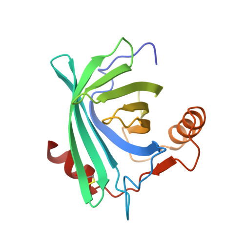
Structure of the epididymal retinoic acid binding protein at 2.1 A resolution.

Newcomer, M.E.

(1993) Structure 1: 7-18

  • DOI: https://doi.org/10.1016/0969-2126(93)90004-z
  • Primary Citation of Related Structures:  
    1EPA, 1EPB

  • PubMed Abstract: 

    Androgen-dependent proteins in the lumen of the epididymis are required for sperm maturation. One of these is a retinoic acid binding protein, E-RABP, which binds both all-trans and 9-cis retinoic acid. The other retinoid-binding proteins whose structures are known do not bind 9-cis retinoids.


  • Organizational Affiliation

    Department of Biochemistry, Vanderbilt University School of Medicine, Nashville, Tennessee 37232-0146.


Macromolecules
Find similar proteins by:  (by identity cutoff)  |  3D Structure
Entity ID: 1
MoleculeChains Sequence LengthOrganismDetailsImage
EPIDIDYMAL RETINOIC ACID-BINDING PROTEIN
A, B
164Rattus norvegicusMutation(s): 0 
Membrane Entity: Yes 
UniProt
Find proteins for P06911 (Rattus norvegicus)
Explore P06911 
Go to UniProtKB:  P06911
Entity Groups  
Sequence Clusters30% Identity50% Identity70% Identity90% Identity95% Identity100% Identity
UniProt GroupP06911
Sequence Annotations
Expand
  • Reference Sequence
Small Molecules
Ligands 1 Unique
IDChains Name / Formula / InChI Key2D Diagram3D Interactions
9CR
Query on 9CR

Download Ideal Coordinates CCD File 
C [auth A],
D [auth B]
(9cis)-retinoic acid
C20 H28 O2
SHGAZHPCJJPHSC-ZVCIMWCZSA-N
Experimental Data & Validation

Experimental Data

  • Method: X-RAY DIFFRACTION
  • Resolution: 2.20 Å
  • R-Value Work: 0.182 
  • R-Value Observed: 0.182 
  • Space Group: P 1 21 1
Unit Cell:
Length ( Å )Angle ( ˚ )
a = 39.8α = 90
b = 58.89β = 109.44
c = 66.65γ = 90
Software Package:
Software NamePurpose
X-PLORmodel building
X-PLORrefinement
X-PLORphasing

Structure Validation

View Full Validation Report


Entry History 

Deposition Data

Revision History  (Full details and data files)

  • Version 1.0: 1994-07-31
    Type: Initial release
  • Version 1.1: 2008-03-24
    Changes: Version format compliance
  • Version 1.2: 2011-07-13
    Changes: Version format compliance
  • Version 1.3: 2016-11-16
    Changes: Non-polymer description
  • Version 1.4: 2017-11-29
    Changes: Derived calculations, Other