1EHI

D-ALANINE:D-LACTATE LIGASE (LMDDL2) OF VANCOMYCIN-RESISTANT LEUCONOSTOC MESENTEROIDES


Experimental Data Snapshot

  • Method: X-RAY DIFFRACTION
  • Resolution: 2.38 Å
  • R-Value Free: 0.257 
  • R-Value Work: 0.184 
  • R-Value Observed: 0.184 

wwPDB Validation   3D Report Full Report


This is version 1.5 of the entry. See complete history


Literature

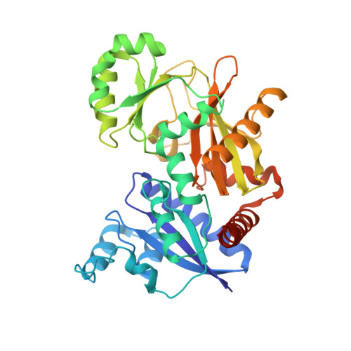
Enzymes of vancomycin resistance: the structure of D-alanine-D-lactate ligase of naturally resistant Leuconostoc mesenteroides.

Kuzin, A.P.Sun, T.Jorczak-Baillass, J.Healy, V.L.Walsh, C.T.Knox, J.R.

(2000) Structure 8: 463-470

  • DOI: https://doi.org/10.1016/s0969-2126(00)00129-5
  • Primary Citation of Related Structures:  
    1EHI

  • PubMed Abstract: 

    The bacterial cell wall and the enzymes that synthesize it are targets of glycopeptide antibiotics (vancomycins and teicoplanins) and beta-lactams (penicillins and cephalosporins). Biosynthesis of cell wall peptidoglycan requires a crosslinking of peptidyl moieties on adjacent glycan strands. The D-alanine-D-alanine transpeptidase, which catalyzes this crosslinking, is the target of beta-lactam antibiotics. Glycopeptides, in contrast, do not inhibit an enzyme, but bind directly to D-alanine-D-alanine and prevent subsequent crosslinking by the transpeptidase. Clinical resistance to vancomycin in enterococcal pathogens has been traced to altered ligases producing D-alanine-D-lactate rather than D-alanine-D-alanine.


  • Organizational Affiliation

    Department of Molecular and Cell Biology, The University of Connecticut, Storrs, CT 06269-3125, USA.


Macromolecules
Find similar proteins by:  (by identity cutoff)  |  3D Structure
Entity ID: 1
MoleculeChains Sequence LengthOrganismDetailsImage
D-ALANINE:D-LACTATE LIGASE
A, B
377Leuconostoc mesenteroidesMutation(s): 0 
EC: 6.3.2.4
UniProt
Find proteins for Q03ZI1 (Leuconostoc mesenteroides subsp. mesenteroides (strain ATCC 8293 / DSM 20343 / BCRC 11652 / CCM 1803 / JCM 6124 / NCDO 523 / NBRC 100496 / NCIMB 8023 / NCTC 12954 / NRRL B-1118 / 37Y))
Explore Q03ZI1 
Go to UniProtKB:  Q03ZI1
Entity Groups  
Sequence Clusters30% Identity50% Identity70% Identity90% Identity95% Identity100% Identity
UniProt GroupQ03ZI1
Sequence Annotations
Expand
  • Reference Sequence
Small Molecules
Ligands 3 Unique
IDChains Name / Formula / InChI Key2D Diagram3D Interactions
ADP
Query on ADP

Download Ideal Coordinates CCD File 
E [auth A]ADENOSINE-5'-DIPHOSPHATE
C10 H15 N5 O10 P2
XTWYTFMLZFPYCI-KQYNXXCUSA-N
PHY
Query on PHY

Download Ideal Coordinates CCD File 
F [auth A]1(S)-AMINOETHYL-(2-CARBOXYPROPYL)PHOSPHORYL-PHOSPHINIC ACID
C6 H15 N O7 P2
BAIYWTZQRMCJBV-DKDXWZAISA-N
MG
Query on MG

Download Ideal Coordinates CCD File 
C [auth A],
D [auth A]
MAGNESIUM ION
Mg
JLVVSXFLKOJNIY-UHFFFAOYSA-N
Experimental Data & Validation

Experimental Data

  • Method: X-RAY DIFFRACTION
  • Resolution: 2.38 Å
  • R-Value Free: 0.257 
  • R-Value Work: 0.184 
  • R-Value Observed: 0.184 
  • Space Group: C 1 2 1
Unit Cell:
Length ( Å )Angle ( ˚ )
a = 118.2α = 90
b = 90.8β = 92.1
c = 82.1γ = 90
Software Package:
Software NamePurpose
SOLVEphasing
X-PLORrefinement
DENZOdata reduction
SCALEPACKdata scaling

Structure Validation

View Full Validation Report


Entry History 

Deposition Data

Revision History  (Full details and data files)

  • Version 1.0: 2000-05-23
    Type: Initial release
  • Version 1.1: 2008-04-27
    Changes: Version format compliance
  • Version 1.2: 2011-07-13
    Changes: Version format compliance
  • Version 1.3: 2017-10-04
    Changes: Refinement description
  • Version 1.4: 2024-02-07
    Changes: Data collection, Database references, Derived calculations
  • Version 1.5: 2024-03-13
    Changes: Source and taxonomy, Structure summary