1EED

X-ray crystallographic analysis of inhibition of endothiapepsin by cyclohexyl renin inhibitors


Experimental Data Snapshot

  • Method: X-RAY DIFFRACTION
  • Resolution: 2.00 Å
  • R-Value Work: 0.152 

wwPDB Validation   3D Report Full Report


This is version 1.4 of the entry. See complete history


Literature

X-ray crystallographic analysis of inhibition of endothiapepsin by cyclohexyl renin inhibitors.

Cooper, J.Quail, W.Frazao, C.Foundling, S.I.Blundell, T.L.Humblet, C.Lunney, E.A.Lowther, W.T.Dunn, B.M.

(1992) Biochemistry 31: 8142-8150

  • DOI: https://doi.org/10.1021/bi00150a005
  • Primary Citation of Related Structures:  
    1EED

  • PubMed Abstract: 

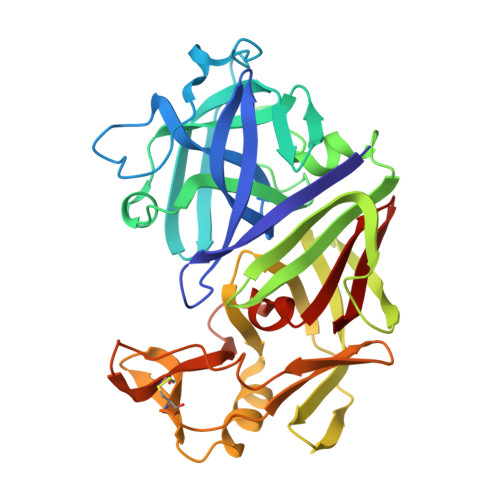
    The crystal structures of endothiapepsin, a fungal aspartic proteinase (EC 3.4.23.6), cocrystallized with two oligopeptide renin inhibitors, PD125967 and PD125754, have been determined at 2.0-A resolution and refined to R-factors of 0.143 and 0.153, respectively. These inhibitors, which are of the hydroxyethylene and statine types, respectively, possess a cyclohexylalanine side chain at P1 and have interesting functionalities at the P3 position which, until now, have not been subjected to crystallographic analysis. PD125967 has a bis(1-naphthylmethyl)acetyl residue at P3, and PD125754 possesses a hydroxyethylene analogue of the P3-P2 peptide bond for proteolytic stability. The structures reveal that the S3 pocket accommodates one naphthyl ring with conformational changes of the Asp 77 and Asp 114 side chains, the other naphthyl group residing in the S4 region. The P3-P2 hydroxyethylene analogue of PD125754 forms a hydrogen bond with the NH of Thr 219, thereby making the same interaction with the enzyme as the equivalent peptide groups of all inhibitors studied so far. The absence of side chains at the P2 and P1' positions of this inhibitor allows water molecules to occupy the respective pockets in the complex. The relative potencies of PD125967 and PD125754 for endothiapepsin are consistent with the changes in solvent-accessible area which take place on inhibitor binding.


  • Organizational Affiliation

    Department of Crystallography, Birkbeck College, London, U.K.


Macromolecules
Find similar proteins by:  (by identity cutoff)  |  3D Structure
Entity ID: 1
MoleculeChains Sequence LengthOrganismDetailsImage
ENDOTHIAPEPSINA [auth P]330Cryphonectria parasiticaMutation(s): 0 
EC: 3.4.23.22
UniProt
Find proteins for P11838 (Cryphonectria parasitica)
Explore P11838 
Go to UniProtKB:  P11838
Entity Groups  
Sequence Clusters30% Identity50% Identity70% Identity90% Identity95% Identity100% Identity
UniProt GroupP11838
Sequence Annotations
Expand
  • Reference Sequence
Small Molecules
Ligands 1 Unique
IDChains Name / Formula / InChI Key2D Diagram3D Interactions
0EO
Query on 0EO

Download Ideal Coordinates CCD File 
B [auth P](2S)-2-[[(3S,4S)-5-cyclohexyl-4-[[(4S,5S)-5-[(2-methylpropan-2-yl)oxycarbonylamino]-4-oxidanyl-6-phenyl-hexanoyl]amino] -3-oxidanyl-pentanoyl]amino]-4-methyl-pentanoic acid
C34 H55 N3 O8
UYUPAZNZXXOTIF-ZIUUJSQJSA-N
Binding Affinity Annotations 
IDSourceBinding Affinity
0EO Binding MOAD:  1EED Ki: 1.62e+4 (nM) from 1 assay(s)
Experimental Data & Validation

Experimental Data

  • Method: X-RAY DIFFRACTION
  • Resolution: 2.00 Å
  • R-Value Work: 0.152 
  • Space Group: P 1 21 1
Unit Cell:
Length ( Å )Angle ( ˚ )
a = 43.1α = 90
b = 75.7β = 96.9
c = 42.9γ = 90
Software Package:
Software NamePurpose
RESTRAINrefinement

Structure Validation

View Full Validation Report


Entry History 

Deposition Data

Revision History  (Full details and data files)

  • Version 1.0: 1994-01-31
    Type: Initial release
  • Version 1.1: 2008-03-03
    Changes: Version format compliance
  • Version 1.2: 2011-07-13
    Changes: Atomic model, Database references, Derived calculations, Non-polymer description, Structure summary, Version format compliance
  • Version 1.3: 2012-12-12
    Changes: Other
  • Version 1.4: 2017-11-29
    Changes: Derived calculations, Other