Chelation of serine 39 to Mg2+ latches a gate at the active site of enolase: structure of the bis(Mg2+) complex of yeast enolase and the intermediate analog phosphonoacetohydroxamate at 2.1-A resolution.
Wedekind, J.E., Poyner, R.R., Reed, G.H., Rayment, I.(1994) Biochemistry 33: 9333-9342
- PubMed: 8049235
- DOI: https://doi.org/10.1021/bi00197a038
- Primary Citation of Related Structures:
1EBG - PubMed Abstract:
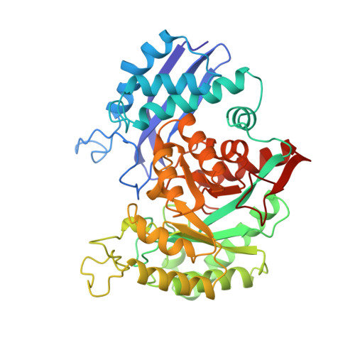
The structure of a new crystal form of enolase from bakers' yeast has been solved to 2.1-A resolution. Crystals were grown from poly(ethylene glycol) and KCl at pH 8.2 in the presence of Mg2+ and a reaction intermediate analog, phosphonoacetohydroxamate (PhAH). Crystals belong to space group C2; have unit cell dimensions a = 123.5 A, b = 73.9 A, and c = 94.8 A with beta = 93.3 degrees; and contain one dimer per asymmetric unit. The structure was solved by molecular replacement from the X-ray coordinates of apoenolase [Stec, B., & Lebioda, L. (1990) J. Mol. Biol. 211, 235-248]. Both essential divalent metal ions are observed to be complexed with the inhibitor. The two Mg2+ ions are 4.05 A apart and are bridged by a mu-oxyl ligand from the carbonyl moiety of PhAH. The "high-affinity" Mg2+ coordinates to the carboxylate side chains of Asp 246, Glu 295, and Asp 320, one water molecule, and the hydroxamate and carbonyl oxygens of PhAH. The second Mg2+ coordinates to a phosphonyl oxygen, two water molecules, and the mu-bridge carbonyl oxygen of PhAH. Coordination schemes with respect to PhAH and water ligands are fully consistent with those of the Mn2+ complexes determined spectroscopically [Poyner, R.R., & Reed, G. H. (1992) Biochemistry 31, 7166-7173]. Remaining ligands for the second Mg2+ are the carbonyl oxygen and gamma-oxygen of Ser 39. Chelation of this Ser residue to Mg2+ effectively "latches" a flexible loop extending from Gly 37 through His 43 and closes off the entrance to the active site. The position of the second Mg2+ in the active site provides new insight into the stereochemistry of substrate binding.
Organizational Affiliation:
Institute for Enzyme Research, Graduate School, University of Wisconsin, Madison 53705.