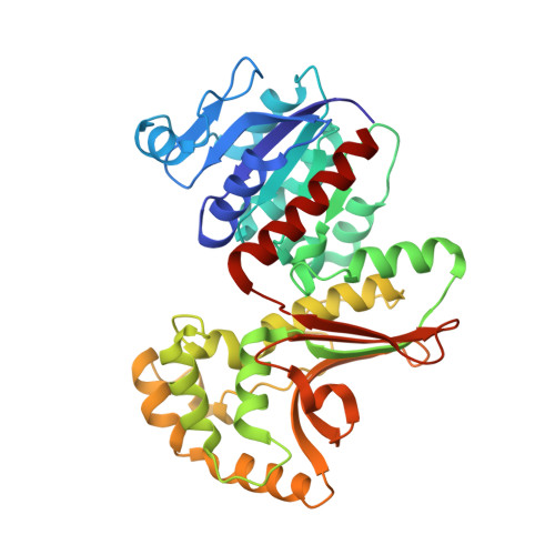
1EBF

HOMOSERINE DEHYDROGENASE FROM S. CEREVISIAE COMPLEX WITH NAD+


Experimental Data Snapshot

  • Method: X-RAY DIFFRACTION
  • Resolution: 2.30 Å
  • R-Value Free: 0.269 
  • R-Value Work: 0.218 
  • R-Value Observed: 0.218 

wwPDB Validation   3D Report Full Report


Ligand Structure Quality Assessment 


This is version 1.4 of the entry. See complete history


Literature

Crystal structures of homoserine dehydrogenase suggest a novel catalytic mechanism for oxidoreductases.

DeLaBarre, B.Thompson, P.R.Wright, G.D.Berghuis, A.M.

(2000) Nat Struct Biol 7: 238-244

  • DOI: https://doi.org/10.1038/73359
  • Primary Citation of Related Structures:  
    1EBF, 1EBU

  • PubMed Abstract: 

    The structure of the antifungal drug target homoserine dehydrogenase (HSD) was determined from Saccharomyces cerevisiae in apo and holo forms, and as a ternary complex with bound products, by X-ray diffraction. The three forms show that the enzyme is a dimer, with each monomer composed of three regions, the nucleotide-binding region, the dimerization region and the catalytic region. The dimerization and catalytic regions have novel folds, whereas the fold of the nucleotide-binding region is a variation on the Rossmann fold. The novel folds impose a novel composition and arrangement of active site residues when compared to all other currently known oxidoreductases. This observation, in conjunction with site-directed mutagenesis of active site residues and steady-state kinetic measurements, suggest that HSD exhibits a new variation on dehydrogenase chemistry.


  • Organizational Affiliation

    Antimicrobial Research Centre and Department of Biochemistry, McMaster University, 1200 Main Street West, Hamilton, Ontario, Canada L8N 3Z5.


Macromolecules
Find similar proteins by:  (by identity cutoff)  |  3D Structure
Entity ID: 1
MoleculeChains Sequence LengthOrganismDetailsImage
HOMOSERINE DEHYDROGENASE
A, B
358Saccharomyces cerevisiaeMutation(s): 0 
Gene Names: HOM6P
EC: 1.1.1.3
UniProt
Find proteins for P31116 (Saccharomyces cerevisiae (strain ATCC 204508 / S288c))
Explore P31116 
Go to UniProtKB:  P31116
Entity Groups  
Sequence Clusters30% Identity50% Identity70% Identity90% Identity95% Identity100% Identity
UniProt GroupP31116
Sequence Annotations
Expand
  • Reference Sequence
Experimental Data & Validation

Experimental Data

  • Method: X-RAY DIFFRACTION
  • Resolution: 2.30 Å
  • R-Value Free: 0.269 
  • R-Value Work: 0.218 
  • R-Value Observed: 0.218 
  • Space Group: P 43 21 2
Unit Cell:
Length ( Å )Angle ( ˚ )
a = 80.396α = 90
b = 80.396β = 90
c = 250.165γ = 90
Software Package:
Software NamePurpose
SOLVEphasing
SHARPphasing
GLRFphasing
CCP4model building
DMmodel building
CNSrefinement
DENZOdata reduction
SCALEPACKdata scaling
CCP4phasing
DMphasing
CNSphasing

Structure Validation

View Full Validation Report


Ligand Structure Quality Assessment 


Entry History 

Deposition Data

Revision History  (Full details and data files)

  • Version 1.0: 2000-03-08
    Type: Initial release
  • Version 1.1: 2008-04-27
    Changes: Version format compliance
  • Version 1.2: 2011-07-13
    Changes: Version format compliance
  • Version 1.3: 2017-10-04
    Changes: Refinement description
  • Version 1.4: 2024-02-07
    Changes: Data collection, Database references, Derived calculations