1EB9

Structure Determinants of Substrate Specificity of Hydroxynitrile Lyase from Manihot esculenta


Experimental Data Snapshot

  • Method: X-RAY DIFFRACTION
  • Resolution: 2.10 Å
  • R-Value Free: 0.244 
  • R-Value Work: 0.197 
  • R-Value Observed: 0.197 

wwPDB Validation   3D Report Full Report


This is version 1.4 of the entry. See complete history


Literature

Structure Determinants of Substrate Specificity of Hydroxynitrile Lyase from Manihot Esculenta.

Lauble, H.Miehlich, B.Foerster, S.Kobler, C.Wajant, H.Effenberger, F.

(2002) Protein Sci 11: 65

  • DOI: https://doi.org/10.1110/ps.33702
  • Primary Citation of Related Structures:  
    1EB8, 1EB9

  • PubMed Abstract: 

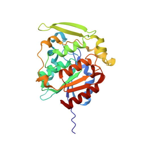
    Tryptophan 128 of hydroxynitrile lyase of Manihot esculenta (MeHNL) covers a significant part of a hydrophobic channel that gives access to the active site of the enzyme. This residue was therefore substituted in the mutant MeHNL-W128A by alanine to study its importance for the substrate specificity of the enzyme. Wild-type MeHNL and MeHNL-W128A showed comparable activity on the natural substrate acetone cyanohydrin (53 and 40 U/mg, respectively). However, the specific activities of MeHNL-W128A for the unnatural substrates mandelonitrile and 4-hydroxymandelonitrile are increased 9-fold and approximately 450-fold, respectively, compared with the wild-type MeHNL. The crystal structure of the MeHNL-W128A substrate-free form at 2.1 A resolution indicates that the W128A substitution has significantly enlarged the active-site channel entrance, and thereby explains the observed changes in substrate specificity for bulky substrates. Surprisingly, the MeHNL-W128A--4-hydroxybenzaldehyde complex structure at 2.1 A resolution shows the presence of two hydroxybenzaldehyde molecules in a sandwich type arrangement in the active site with an additional hydrogen bridge to the reacting center.


  • Organizational Affiliation

    Institut für Organische Chemie der Universität Stuttgart, D-70569 Stuttgart, Germany. PeterLauble@t-online.de


Macromolecules
Find similar proteins by:  (by identity cutoff)  |  3D Structure
Entity ID: 1
MoleculeChains Sequence LengthOrganismDetailsImage
HYDROXYNITRILE LYASE
A, B
262Manihot esculentaMutation(s): 1 
EC: 4.2.1.37
UniProt
Find proteins for P52705 (Manihot esculenta)
Explore P52705 
Go to UniProtKB:  P52705
Entity Groups  
Sequence Clusters30% Identity50% Identity70% Identity90% Identity95% Identity100% Identity
UniProt GroupP52705
Sequence Annotations
Expand
  • Reference Sequence
Experimental Data & Validation

Experimental Data

  • Method: X-RAY DIFFRACTION
  • Resolution: 2.10 Å
  • R-Value Free: 0.244 
  • R-Value Work: 0.197 
  • R-Value Observed: 0.197 
  • Space Group: P 41 21 2
Unit Cell:
Length ( Å )Angle ( ˚ )
a = 106.34α = 90
b = 106.34β = 90
c = 188.84γ = 90
Software Package:
Software NamePurpose
X-PLORrefinement
DENZOdata reduction
SCALEPACKdata scaling
AMoREphasing

Structure Validation

View Full Validation Report


Entry History 

Deposition Data

Revision History  (Full details and data files)

  • Version 1.0: 2002-01-01
    Type: Initial release
  • Version 1.1: 2011-05-08
    Changes: Version format compliance
  • Version 1.2: 2011-07-13
    Changes: Version format compliance
  • Version 1.3: 2019-07-24
    Changes: Data collection
  • Version 1.4: 2023-12-13
    Changes: Data collection, Database references, Other, Refinement description