1E7J

HMG-D complexed to a bulge DNA


Experimental Data Snapshot

  • Method: SOLUTION NMR
  • Conformers Calculated: 50 
  • Conformers Submitted: 10 
  • Selection Criteria: LOW VALUES OF THE NOE ENERGY TERM AND PROTEIN LOCATED WITHIN MINOR GROOVE OF DNA 

wwPDB Validation   3D Report Full Report


This is version 1.2 of the entry. See complete history


Literature

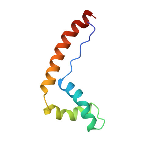
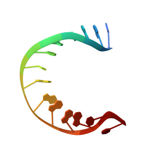
Hmg-D Complexed to a Bulge DNA: An NMR Model

Cerdan, R.Payet, D.Yang, J.-C.Travers, A.A.Neuhaus, D.

(2001) Protein Sci 10: 504

  • DOI: https://doi.org/10.1110/ps.35501
  • Primary Citation of Related Structures:  
    1E7J

  • PubMed Abstract: 

    An NMR model is presented for the structure of HMG-D, one of the Drosophila counterparts of mammalian HMG1/2 proteins, bound to a particular distorted DNA structure, a dA(2) DNA bulge. The complex is in fast to intermediate exchange on the NMR chemical shift time scale and suffers substantial linebroadening for the majority of interfacial resonances. This essentially precludes determination of a high-resolution structure for the interface based on NMR data alone. However, by introducing a small number of additional constraints based on chemical shift and linewidth footprinting combined with analogies to known structures, an ensemble of model structures was generated using a computational strategy equivalent to that for a conventional NMR structure determination. We find that the base pair adjacent to the dA(2) bulge is not formed and that the protein recognizes this feature in forming the complex; intermolecular NOE enhancements are observed from the sidechain of Thr 33 to all four nucleotides of the DNA sequence step adjacent to the bulge. Our results form the first experimental demonstration that when binding to deformed DNA, non-sequence-specific HMG proteins recognize the junction between duplex and nonduplex DNA. Similarities and differences of the present structural model relative to other HMG-DNA complex structures are discussed.


  • Organizational Affiliation

    M.R.C. Laboratory of Molecular Biology, Cambridge CB2 2QH, UK.


Macromolecules

Find similar proteins by:  (by identity cutoff)  |  3D Structure
Entity ID: 1
MoleculeChains Sequence LengthOrganismDetailsImage
HIGH MOBILITY GROUP PROTEIN D74Drosophila melanogasterMutation(s): 0 
UniProt
Find proteins for Q05783 (Drosophila melanogaster)
Explore Q05783 
Go to UniProtKB:  Q05783
Entity Groups  
Sequence Clusters30% Identity50% Identity70% Identity90% Identity95% Identity100% Identity
UniProt GroupQ05783
Sequence Annotations
Expand
  • Reference Sequence

Find similar nucleic acids by:  Sequence   |   3D Structure  

Entity ID: 2
MoleculeChains LengthOrganismImage
DNA (5'-D(*CP*GP*AP*TP*AP*TP*TP*AP*AP*GP*AP*GP*CP*C)-3')14N/A
Sequence Annotations
Expand
  • Reference Sequence

Find similar nucleic acids by:  Sequence   |   3D Structure  

Entity ID: 3
MoleculeChains LengthOrganismImage
DNA (5'-D(*GP*GP*CP*TP*CP*AP*AP*TP*AP*TP*CP*G)-3')12N/A
Sequence Annotations
Expand
  • Reference Sequence
Small Molecules
Binding Affinity Annotations 
IDSourceBinding Affinity
DNA PDBBind:  1E7J Kd: 1000 (nM) from 1 assay(s)
Experimental Data & Validation

Experimental Data

  • Method: SOLUTION NMR
  • Conformers Calculated: 50 
  • Conformers Submitted: 10 
  • Selection Criteria: LOW VALUES OF THE NOE ENERGY TERM AND PROTEIN LOCATED WITHIN MINOR GROOVE OF DNA 

Structure Validation

View Full Validation Report


Entry History 

Deposition Data

Revision History  (Full details and data files)

  • Version 1.0: 2001-03-05
    Type: Initial release
  • Version 1.1: 2011-05-08
    Changes: Version format compliance
  • Version 1.2: 2011-07-13
    Changes: Version format compliance