Mechanistic Implications for Escherichia Coli Cofactor-Dependent Phosphoglycerate Mutase Based on the High-Resolution Crystal Structure of a Vanadate Complex.
Bond, C.S., White, M., Hunter, W.N.(2002) J Mol Biol 316: 1071
- PubMed: 11884145
- DOI: https://doi.org/10.1006/jmbi.2002.5418
- Primary Citation of Related Structures:
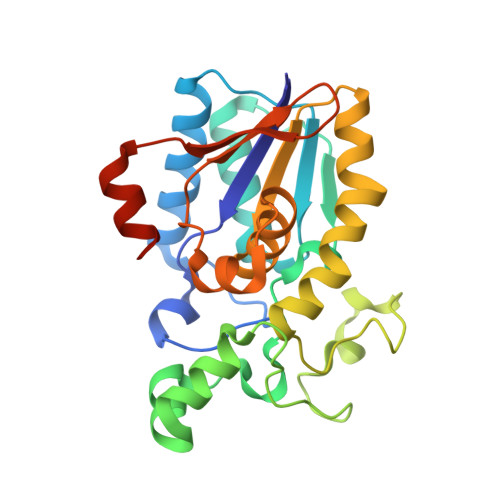
1E59 - PubMed Abstract:
The structure of Escherichia coli cofactor-dependent phosphoglycerate mutase (dPGM), complexed with the potent inhibitor vanadate, has been determined to a resolution of 1.30 A (R-factor 0.159; R-free 0.213). The inhibitor is present in the active site, principally as divanadate, but with evidence of additional vanadate moieties at either end, and representing a different binding mode to that observed in the structural homologue prostatic acid phosphatase. The analysis reveals the enzyme-ligand interactions involved in inhibition of the mutase activity by vanadate and identifies a water molecule, observed in the native E.coli dPGM structure which, once activated by vanadate, may dephosphorylate the active protein. Rather than reflecting the active conformation previously observed for E.coli dPGM, the inhibited protein's conformation resembles that of the inactive dephosphorylated Saccharomyces cerevisiae dPGM. The provision of a high-resolution structure of both active and inactive forms of dPGM from a single organism, in conjunction with computational modelling of substrate molecules in the active site provides insight into the binding of substrates and the specific interactions necessary for three different activities, mutase, synthase and phosphatase, within a single active site. The sequence similarity of E.coli and human dPGMs allows us to correlate structure with clinical pathology.
Organizational Affiliation:
Division of Biological Chemistry and Molecular Microbiology, Wellcome Trust Biocentre, University of Dundee, Dundee DD1 5EH, Scotland, UK.