1E58

E.coli cofactor-dependent phosphoglycerate mutase


Experimental Data Snapshot

  • Method: X-RAY DIFFRACTION
  • Resolution: 1.25 Å
  • R-Value Free: 0.168 
  • R-Value Observed: 0.121 

wwPDB Validation   3D Report Full Report


This is version 1.3 of the entry. See complete history


Literature

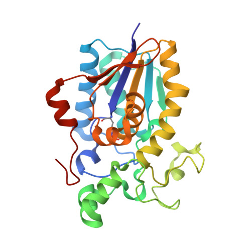
High Resolution Structure of the Phosphohistidine-Activated Form of Escherichia Coli Cofactor-Dependent Phosphoglycerate Mutase.

Bond, C.S.White, M.F.Hunter, W.N.

(2001) J Biol Chem 276: 3247

  • DOI: https://doi.org/10.1074/jbc.M007318200
  • Primary Citation of Related Structures:  
    1E58

  • PubMed Abstract: 

    The active conformation of the dimeric cofactor-dependent phosphoglycerate mutase (dPGM) from Escherichia coli has been elucidated by crystallographic methods to a resolution of 1.25 A (R-factor 0.121; R-free 0.168). The active site residue His(10), central in the catalytic mechanism of dPGM, is present as a phosphohistidine with occupancy of 0.28. The structural changes on histidine phosphorylation highlight various features that are significant in the catalytic mechanism. The C-terminal 10-residue tail, which is not observed in previous dPGM structures, is well ordered and interacts with residues implicated in substrate binding; the displacement of a loop adjacent to the active histidine brings previously overlooked residues into positions where they may directly influence catalysis. E. coli dPGM, like the mammalian dPGMs, is a dimer, whereas previous structural work has concentrated on monomeric and tetrameric yeast forms. We can now analyze the sequence differences that cause this variation of quaternary structure.


  • Organizational Affiliation

    Wellcome Trust Biocentre, University of Dundee, Dundee DD1 5EH, United Kingdom.


Macromolecules
Find similar proteins by:  (by identity cutoff)  |  3D Structure
Entity ID: 1
MoleculeChains Sequence LengthOrganismDetailsImage
PHOSPHOGLYCERATE MUTASE249Escherichia coli K-12Mutation(s): 0 
Gene Names: PGM1
EC: 5.4.2.1
UniProt
Find proteins for P62707 (Escherichia coli (strain K12))
Explore P62707 
Go to UniProtKB:  P62707
Entity Groups  
Sequence Clusters30% Identity50% Identity70% Identity90% Identity95% Identity100% Identity
UniProt GroupP62707
Sequence Annotations
Expand
  • Reference Sequence
Small Molecules
Modified Residues  1 Unique
IDChains TypeFormula2D DiagramParent
NEP
Query on NEP
A
L-PEPTIDE LINKINGC6 H10 N3 O5 PHIS
Experimental Data & Validation

Experimental Data

  • Method: X-RAY DIFFRACTION
  • Resolution: 1.25 Å
  • R-Value Free: 0.168 
  • R-Value Observed: 0.121 
  • Space Group: P 21 21 2
Unit Cell:
Length ( Å )Angle ( ˚ )
a = 61.57α = 90
b = 113β = 90
c = 40.26γ = 90
Software Package:
Software NamePurpose
SHELXL-97refinement
DENZOdata reduction
SCALEPACKdata scaling
SHELXphasing

Structure Validation

View Full Validation Report


Entry History 

Deposition Data

Revision History  (Full details and data files)

  • Version 1.0: 2001-03-20
    Type: Initial release
  • Version 1.1: 2011-05-08
    Changes: Version format compliance
  • Version 1.2: 2011-07-13
    Changes: Version format compliance
  • Version 1.3: 2019-05-22
    Changes: Data collection, Derived calculations, Other, Refinement description