Crystal Structure of Nadp(H)-Dependent Ketose Reductase from Besimia Argentifolii at 2.3 Angstrom Resolution
Banfield, M.J., Salvucci, M.E., Baker, E.N., Smith, C.A.(2001) J Mol Biol 306: 239
- PubMed: 11237597
- DOI: https://doi.org/10.1006/jmbi.2000.4381
- Primary Citation of Related Structures:
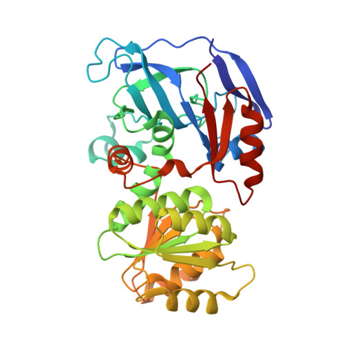
1E3J - PubMed Abstract:
Polyhydric alcohols are widely found in nature and can be accumulated to high concentrations as a protection against a variety of environmental stresses. It is only recently, however, that these molecules have been shown to be active in protection against heat stress, specifically in the use of sorbitol by the silverleaf whitefly, Bemisia argentifolii. We have determined the structure of the enzyme responsible for production of sorbitol in Bemisia argentifolii, NADP(H)-dependent ketose reductase (BaKR), to 2.3 A resolution. The structure was solved by multiwavelength anomalous diffraction (MAD) using the anomalous scattering from two zinc atoms bound in the structure, and was refined to an R factor of 21.9 % (R(free)=25.1 %). BaKR belongs to the medium-chain dehydrogenase family and its structure is the first for the sorbitol dehydrogenase branch of this family. The enzyme is tetrameric, with the monomer having a very similar fold to the alcohol dehydrogenases (ADHs). Although the structure determined is for the apo form, a phosphate ion in the active site marks the likely position for the adenyl phosphate of NADP(H). The catalytic zinc ion is tetrahedrally coordinated to Cys41, His66, Glu67 and a water molecule, in a modification of the zinc site usually found in ADHs. This modified zinc site seems likely to be a conserved feature of the sorbitol dehydrogenase sub-family. Comparisons with other members of the ADH family have also enabled us to model a ternary complex of the enzyme, and suggest how structural differences may influence coenzyme binding and substrate specificity in the reduction of fructose to sorbitol.
Organizational Affiliation:
School of Biological Sciences, University of Auckland, Private Bag 92-019, Auckland, New Zealand.