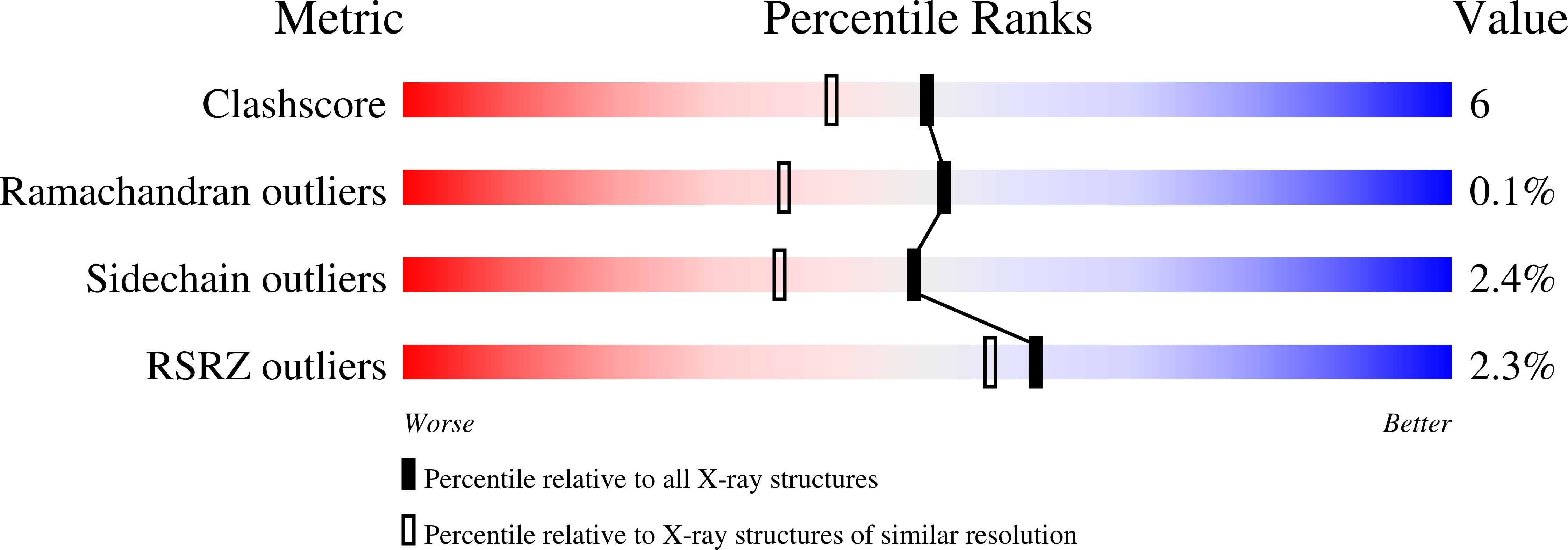
1E3A

A slow processing precursor penicillin acylase from Escherichia coli


Experimental Data Snapshot

  • Method: X-RAY DIFFRACTION
  • Resolution: 1.80 Å
  • R-Value Free: 0.197 
  • R-Value Work: 0.158 
  • R-Value Observed: 0.149 

wwPDB Validation   3D Report Full Report


This is version 1.3 of the entry. See complete history


Literature

Structure of a Slow Processing Precursor Penicillin Acylase from Escherichia Coli Reveals the Linker Peptide Blocking the Active-Site Cleft

Hewitt, L.Kasche, V.Lummer, K.Lewis, R.J.Murshudov, G.N.Verma, C.S.Dodson, G.G.Wilson, K.S.

(2000) J Mol Biol 302: 887

  • DOI: https://doi.org/10.1006/jmbi.2000.4105
  • Primary Citation of Related Structures:  
    1E3A

  • PubMed Abstract: 

    Penicillin G acylase is a periplasmic protein, cytoplasmically expressed as a precursor polypeptide comprising a signal sequence, the A and B chains of the mature enzyme (209 and 557 residues respectively) joined by a spacer peptide of 54 amino acid residues. The wild-type AB heterodimer is produced by proteolytic removal of this spacer in the periplasm. The first step in processing is believed to be autocatalytic hydrolysis of the peptide bond between the C-terminal residue of the spacer and the active-site serine residue at the N terminus of the B chain. We have determined the crystal structure of a slowly processing precursor mutant (Thr263Gly) of penicillin G acylase from Escherichia coli, which reveals that the spacer peptide blocks the entrance to the active-site cleft consistent with an autocatalytic mechanism of maturation. In this mutant precursor there is, however, an unexpected cleavage at a site four residues from the active-site serine residue. Analyses of the stereochemistry of the 260-261 bond seen to be cleaved in this precursor structure and of the 263-264 peptide bond have suggested factors that may govern the autocatalytic mechanism.


  • Organizational Affiliation

    Department of Chemistry, University of York, Heslington York, YO10 5DD, UK.


Macromolecules
Find similar proteins by:  (by identity cutoff)  |  3D Structure
Entity ID: 1
MoleculeChains Sequence LengthOrganismDetailsImage
PENICILLIN AMIDASE ALPHA SUBUNIT260Escherichia coliMutation(s): 0 
Gene Names: PAC
EC: 3.5.1.11
UniProt
Find proteins for P06875 (Escherichia coli)
Explore P06875 
Go to UniProtKB:  P06875
Entity Groups  
Sequence Clusters30% Identity50% Identity70% Identity90% Identity95% Identity100% Identity
UniProt GroupP06875
Sequence Annotations
Expand
  • Reference Sequence
Find similar proteins by:  (by identity cutoff)  |  3D Structure
Entity ID: 2
MoleculeChains Sequence LengthOrganismDetailsImage
PENICILLIN AMIDASE BETA SUBUNIT560Escherichia coliMutation(s): 1 
Gene Names: PAC
EC: 3.5.1.11
UniProt
Find proteins for P06875 (Escherichia coli)
Explore P06875 
Go to UniProtKB:  P06875
Entity Groups  
Sequence Clusters30% Identity50% Identity70% Identity90% Identity95% Identity100% Identity
UniProt GroupP06875
Sequence Annotations
Expand
  • Reference Sequence
Experimental Data & Validation

Experimental Data

  • Method: X-RAY DIFFRACTION
  • Resolution: 1.80 Å
  • R-Value Free: 0.197 
  • R-Value Work: 0.158 
  • R-Value Observed: 0.149 
  • Space Group: P 1
Unit Cell:
Length ( Å )Angle ( ˚ )
a = 50.71α = 66.14
b = 64.27β = 74.18
c = 72γ = 74.23
Software Package:
Software NamePurpose
REFMACrefinement
DENZOdata reduction
SCALEPACKdata scaling
AMoREphasing

Structure Validation

View Full Validation Report


Entry History 

Deposition Data

Revision History  (Full details and data files)

  • Version 1.0: 2000-11-29
    Type: Initial release
  • Version 1.1: 2014-10-22
    Changes: Data collection, Database references, Derived calculations, Other, Version format compliance
  • Version 1.2: 2019-07-24
    Changes: Data collection
  • Version 1.3: 2023-12-13
    Changes: Data collection, Database references, Derived calculations, Refinement description