1DYR

THE STRUCTURE OF PNEUMOCYSTIS CARINII DIHYDROFOLATE REDUCTASE TO 1.9 ANGSTROMS RESOLUTION


Experimental Data Snapshot

  • Method: X-RAY DIFFRACTION
  • Resolution: 1.86 Å
  • R-Value Observed: 0.181 

wwPDB Validation   3D Report Full Report


This is version 1.3 of the entry. See complete history


Literature

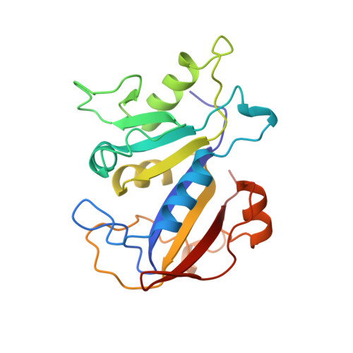
The structure of Pneumocystis carinii dihydrofolate reductase to 1.9 A resolution.

Champness, J.N.Achari, A.Ballantine, S.P.Bryant, P.K.Delves, C.J.Stammers, D.K.

(1994) Structure 2: 915-924

  • DOI: https://doi.org/10.1016/s0969-2126(94)00093-x
  • Primary Citation of Related Structures:  
    1DYR

  • PubMed Abstract: 

    The fungal pathogen Pneumocystis carinii causes a pneumonia which is an opportunistic infection of AIDS patients. Current therapy includes the dihydrofolate reductase (DHFR) inhibitor trimethoprim which is selective but only a relatively weak inhibitor of the enzyme for P. carinii. Determination of the three-dimensional structure of the enzyme should form the basis for design of more potent and selective therapeutic agents for treatment of the disease.


  • Organizational Affiliation

    Physical Sciences Department, Wellcome Research Laboratories, Beckenham, Kent, UK.


Macromolecules
Find similar proteins by:  (by identity cutoff)  |  3D Structure
Entity ID: 1
MoleculeChains Sequence LengthOrganismDetailsImage
DIHYDROFOLATE REDUCTASE206Pneumocystis cariniiMutation(s): 0 
Gene Names: C-DNA P.CARINII DHFR
EC: 1.5.1.3
UniProt
Find proteins for P16184 (Pneumocystis carinii)
Explore P16184 
Go to UniProtKB:  P16184
Entity Groups  
Sequence Clusters30% Identity50% Identity70% Identity90% Identity95% Identity100% Identity
UniProt GroupP16184
Sequence Annotations
Expand
  • Reference Sequence
Small Molecules
Ligands 2 Unique
IDChains Name / Formula / InChI Key2D Diagram3D Interactions
NDP
Query on NDP

Download Ideal Coordinates CCD File 
B [auth A]NADPH DIHYDRO-NICOTINAMIDE-ADENINE-DINUCLEOTIDE PHOSPHATE
C21 H30 N7 O17 P3
ACFIXJIJDZMPPO-NNYOXOHSSA-N
TOP
Query on TOP

Download Ideal Coordinates CCD File 
C [auth A]TRIMETHOPRIM
C14 H18 N4 O3
IEDVJHCEMCRBQM-UHFFFAOYSA-N
Binding Affinity Annotations 
IDSourceBinding Affinity
TOP BindingDB:  1DYR Ki: min: 151.36, max: 2.80e+5 (nM) from 3 assay(s)
IC50: min: 1.20e+4, max: 4.30e+4 (nM) from 6 assay(s)
Binding MOAD:  1DYR IC50: 2.00e+4 (nM) from 1 assay(s)
Experimental Data & Validation

Experimental Data

  • Method: X-RAY DIFFRACTION
  • Resolution: 1.86 Å
  • R-Value Observed: 0.181 
  • Space Group: P 1 21 1
Unit Cell:
Length ( Å )Angle ( ˚ )
a = 69.9α = 90
b = 43.6β = 117.7
c = 37.6γ = 90
Software Package:
Software NamePurpose
PROFFTrefinement
XENGENdata reduction

Structure Validation

View Full Validation Report


Entry History 

Deposition Data

Revision History  (Full details and data files)

  • Version 1.0: 1995-10-15
    Type: Initial release
  • Version 1.1: 2008-03-03
    Changes: Version format compliance
  • Version 1.2: 2011-07-13
    Changes: Version format compliance
  • Version 1.3: 2024-02-07
    Changes: Data collection, Database references, Derived calculations, Other, Structure summary