1DYK

Laminin alpha 2 chain LG4-5 domain pair


Experimental Data Snapshot

  • Method: X-RAY DIFFRACTION
  • Resolution: 2.00 Å
  • R-Value Free: 0.261 
  • R-Value Work: 0.233 
  • R-Value Observed: 0.233 

wwPDB Validation   3D Report Full Report


This is version 1.4 of the entry. See complete history


Literature

Structure of the C-Terminal Laminin G-Like Domain Pair of the Laminin Alpha 2 Chain Harbouring Binding Sites for Alpha-Dystroglycan and Heparin

Tisi, D.Talts, J.F.Timpl, R.Hohenester, E.

(2000) EMBO J 19: 1432

  • DOI: https://doi.org/10.1093/emboj/19.7.1432
  • Primary Citation of Related Structures:  
    1DYK

  • PubMed Abstract: 

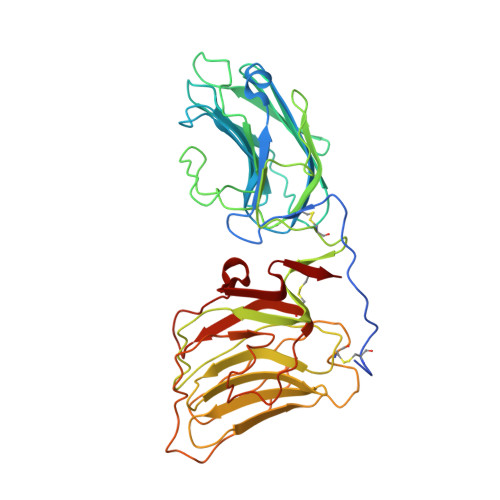
    The laminins are large heterotrimeric glycoproteins with fundamental roles in basement membrane architecture and function. The C-terminus of the laminin alpha chain contains a tandem of five laminin G-like (LG) domains. We report the 2.0 A crystal structure of the laminin alpha2 LG4-LG5 domain pair, which harbours binding sites for heparin and the cell surface receptor alpha-dystroglycan, and is 41% identical to the laminin alpha1 E3 fragment. LG4 and LG5 are arranged in a V-shaped fashion related by a 110 degrees rotation about an axis passing near the domain termini. An extended N-terminal segment is disulfide bonded to LG5 and stabilizes the domain pair. Two calcium ions, one each in LG4 and LG5, are located 65 A apart at the tips of the domains opposite the polypeptide termini. An extensive basic surface region between the calcium sites is proposed to bind alpha-dystroglycan and heparin. The LG4-LG5 structure was used to construct a model of the laminin LG1-LG5 tandem and interpret missense mutations underlying protein S deficiency.


  • Organizational Affiliation

    Abteilung Proteinchemie, Max-Planck-Institut für Biochemie, D-82152 Martinsried, Germany.


Macromolecules
Find similar proteins by:  (by identity cutoff)  |  3D Structure
Entity ID: 1
MoleculeChains Sequence LengthOrganismDetailsImage
LAMININ ALPHA 2 CHAIN394Mus musculusMutation(s): 0 
UniProt
Find proteins for Q60675 (Mus musculus)
Explore Q60675 
Go to UniProtKB:  Q60675
Entity Groups  
Sequence Clusters30% Identity50% Identity70% Identity90% Identity95% Identity100% Identity
UniProt GroupQ60675
Sequence Annotations
Expand
  • Reference Sequence
Experimental Data & Validation

Experimental Data

  • Method: X-RAY DIFFRACTION
  • Resolution: 2.00 Å
  • R-Value Free: 0.261 
  • R-Value Work: 0.233 
  • R-Value Observed: 0.233 
  • Space Group: C 2 2 21
Unit Cell:
Length ( Å )Angle ( ˚ )
a = 70.6α = 90
b = 111.5β = 90
c = 124.8γ = 90
Software Package:
Software NamePurpose
X-PLORrefinement
MOSFLMdata reduction
SCALAdata scaling
X-PLORphasing

Structure Validation

View Full Validation Report


Entry History 

Deposition Data

Revision History  (Full details and data files)

  • Version 1.0: 2001-02-04
    Type: Initial release
  • Version 1.1: 2011-05-07
    Changes: Version format compliance
  • Version 1.2: 2011-07-13
    Changes: Version format compliance
  • Version 1.3: 2019-05-22
    Changes: Data collection, Other, Refinement description
  • Version 1.4: 2023-12-06
    Changes: Data collection, Database references, Derived calculations, Other, Refinement description