1DTJ

CRYSTAL STRUCTURE OF NOVA-2 KH3 K-HOMOLOGY RNA-BINDING DOMAIN


Experimental Data Snapshot

  • Method: X-RAY DIFFRACTION
  • Resolution: 2.00 Å
  • R-Value Free: 0.279 
  • R-Value Work: 0.225 
  • R-Value Observed: 0.229 

wwPDB Validation   3D Report Full Report


This is version 1.5 of the entry. See complete history


Literature

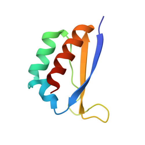
Crystal structures of Nova-1 and Nova-2 K-homology RNA-binding domains.

Lewis, H.A.Chen, H.Edo, C.Buckanovich, R.J.Yang, Y.Y.Musunuru, K.Zhong, R.Darnell, R.B.Burley, S.K.

(1999) Structure 7: 191-203

  • DOI: https://doi.org/10.1016/S0969-2126(99)80025-2
  • Primary Citation of Related Structures:  
    1DT4, 1DTJ

  • PubMed Abstract: 

    Nova-1 and Nova-2 are related neuronal proteins that were initially cloned using antisera obtained from patients with the autoimmune neurological disease paraneoplastic opsoclonus-myoclonus ataxia (POMA). Both of these disease gene products contain three RNA-binding motifs known as K-homology or KH domains, and their RNA ligands have been identified via binding-site selection experiments. The KH motif structure has been determined previously using NMR spectroscopy, but not using X-ray crystallography. Many proteins contain more than one KH domain, yet there is no published structural information regarding the behavior of such multimers.


  • Organizational Affiliation

    Laboratories of Molecular Biophysics, Howard Hughes Medical Institute, The Rockefeller University, 1230 York Avenue, New York, NY, 10021 USA.


Macromolecules
Find similar proteins by:  (by identity cutoff)  |  3D Structure
Entity ID: 1
MoleculeChains Sequence LengthOrganismDetailsImage
RNA-BINDING NEUROONCOLOGICAL VENTRAL ANTIGEN 2
A, B, C, D
76Homo sapiensMutation(s): 1 
UniProt & NIH Common Fund Data Resources
Find proteins for Q9UNW9 (Homo sapiens)
Explore Q9UNW9 
Go to UniProtKB:  Q9UNW9
PHAROS:  Q9UNW9
GTEx:  ENSG00000104967 
Entity Groups  
Sequence Clusters30% Identity50% Identity70% Identity90% Identity95% Identity100% Identity
UniProt GroupQ9UNW9
Sequence Annotations
Expand
  • Reference Sequence
Experimental Data & Validation

Experimental Data

  • Method: X-RAY DIFFRACTION
  • Resolution: 2.00 Å
  • R-Value Free: 0.279 
  • R-Value Work: 0.225 
  • R-Value Observed: 0.229 
  • Space Group: C 1 2 1
Unit Cell:
Length ( Å )Angle ( ˚ )
a = 66.5α = 90
b = 61.1β = 98.2
c = 97.3γ = 90
Software Package:
Software NamePurpose
CNSrefinement
DENZOdata reduction
CCP4data scaling
CNSphasing

Structure Validation

View Full Validation Report


Entry History 

Deposition Data

Revision History  (Full details and data files)

  • Version 1.0: 2000-02-18
    Type: Initial release
  • Version 1.1: 2008-04-27
    Changes: Version format compliance
  • Version 1.2: 2011-07-13
    Changes: Version format compliance
  • Version 1.3: 2021-02-03
    Changes: Database references, Structure summary
  • Version 1.4: 2021-11-03
    Changes: Database references, Derived calculations
  • Version 1.5: 2024-02-07
    Changes: Data collection