1DOS

STRUCTURE OF FRUCTOSE-BISPHOSPHATE ALDOLASE


Experimental Data Snapshot

  • Method: X-RAY DIFFRACTION
  • Resolution: 1.67 Å
  • R-Value Free: 0.204 
  • R-Value Work: 0.171 
  • R-Value Observed: 0.171 

wwPDB Validation   3D Report Full Report


This is version 1.3 of the entry. See complete history


Literature

Novel active site in Escherichia coli fructose 1,6-bisphosphate aldolase.

Blom, N.S.Tetreault, S.Coulombe, R.Sygusch, J.

(1996) Nat Struct Biol 3: 856-862

  • DOI: https://doi.org/10.1038/nsb1096-856
  • Primary Citation of Related Structures:  
    1DOS

  • PubMed Abstract: 

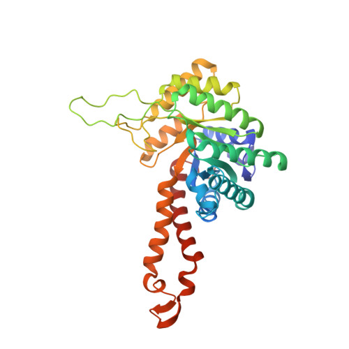
    The molecular architecture of the Class II E. coli fructose 1,6-bisphosphate aldolase dimer was determined to 1.6 A resolution. The subunit fold corresponds to a singly wound alpha/beta-barrel with an active site located on the beta-barrel carboxyl side of each subunit. In each subunit there are two mutually exclusive zinc metal ion binding sites, 3.2 A apart; the exclusivity is mediated by a conformational transition involving side-chain rotations by chelating histidine residues. A binding site for K+ and NH4+ activators was found near the beta-barrel centre. Although Class I and Class II aldolases catalyse identical reactions, their active sites do not share common amino acid residues, are structurally dissimilar, and from sequence comparisons appear to be evolutionary distinct.


  • Organizational Affiliation

    Départment de biochimie, Université de Montréal, Canada.


Macromolecules
Find similar proteins by:  (by identity cutoff)  |  3D Structure
Entity ID: 1
MoleculeChains Sequence LengthOrganismDetailsImage
ALDOLASE CLASS II
A, B
358Escherichia coliMutation(s): 0 
EC: 4.1.2.13
UniProt
Find proteins for P0AB71 (Escherichia coli (strain K12))
Explore P0AB71 
Go to UniProtKB:  P0AB71
Entity Groups  
Sequence Clusters30% Identity50% Identity70% Identity90% Identity95% Identity100% Identity
UniProt GroupP0AB71
Sequence Annotations
Expand
  • Reference Sequence
Experimental Data & Validation

Experimental Data

  • Method: X-RAY DIFFRACTION
  • Resolution: 1.67 Å
  • R-Value Free: 0.204 
  • R-Value Work: 0.171 
  • R-Value Observed: 0.171 
  • Space Group: P 1 21 1
Unit Cell:
Length ( Å )Angle ( ˚ )
a = 90.53α = 90
b = 73.38β = 106.6
c = 57.8γ = 90
Software Package:
Software NamePurpose
X-PLORmodel building
X-PLORrefinement
SDMSdata reduction
X-PLORphasing

Structure Validation

View Full Validation Report


Entry History 

Deposition Data

Revision History  (Full details and data files)

  • Version 1.0: 1997-07-07
    Type: Initial release
  • Version 1.1: 2008-03-03
    Changes: Version format compliance
  • Version 1.2: 2011-07-13
    Changes: Version format compliance
  • Version 1.3: 2024-02-07
    Changes: Data collection, Database references, Derived calculations, Other