1DLK

CRYSTAL STRUCTURE ANALYSIS OF DELTA-CHYMOTRYPSIN BOUND TO A PEPTIDYL CHLOROMETHYL KETONE INHIBITOR


Experimental Data Snapshot

  • Method: X-RAY DIFFRACTION
  • Resolution: 2.14 Å
  • R-Value Free: 0.245 
  • R-Value Work: 0.207 
  • R-Value Observed: 0.206 

wwPDB Validation   3D Report Full Report


This is version 1.3 of the entry. See complete history


Literature

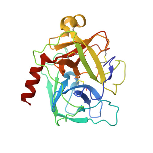
Crystal structure of delta-chymotrypsin bound to a peptidyl chloromethyl ketone inhibitor.

Mac Sweeney, A.Birrane, G.Walsh, M.A.O'Connell, T.Malthouse, J.P.Higgins, T.M.

(2000) Acta Crystallogr D Biol Crystallogr 56: 280-286

  • DOI: https://doi.org/10.1107/s0907444999016583
  • Primary Citation of Related Structures:  
    1DLK

  • PubMed Abstract: 

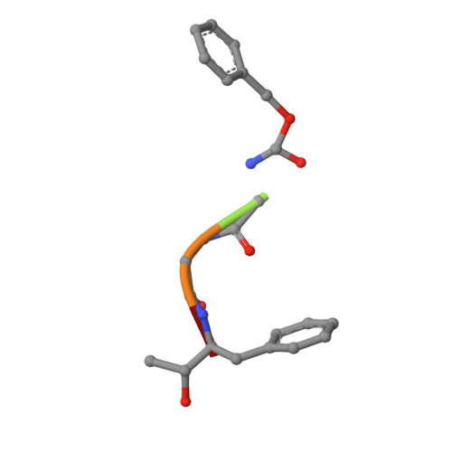
    Chymotrypsin is a member of the trypsin family of serine proteases and is one of the first proteins successfully studied by X-ray crystallography. It is secreted into the intestine as the inactive precursor chymotrypsinogen; four sequential cleavages of the peptide bonds following residues 13, 15, 146 and 148 occur to generate the active pi, delta, kappa and alpha forms of chymotrypsin. (13)C NMR has shown [O'Connell & Malthouse (1995). Biochem. J. 307, 353-359] that when the delta form of chymotrypsin is inhibited by 2-(13)C-enriched benzyloxycarbonylglycylglycylphenylalanyl chloromethane, a tetrahedral adduct is formed which is thought to be analogous to the tetrahedral intermediate formed during catalysis. This inhibitor complex has been crystallized as a dimer in space group P4(1)2(1)2. The structure has been refined at 2.14 A resolution to an R value of 21.2% (free R = 25.2%). Conformational differences between delta-chymotrypsin and chymotrypsinogen in the region of the flexible autolysis loop (residues 145-150) were observed. This is the first crystal structure of delta-chymotrypsin and includes two residues which are disordered in previous crystal structures of active chymotrypsin. A difference of 11.3 A(2) between the average B values of the monomers within the asymmetric unit is caused by lattice-disordering effects approximating to rotation of the molecules about a crystallographic screw axis. The substrate-binding mode of the inhibitor was similar to other chymotrypsin peptidyl inhibitor complexes, but this is the first published chymotrypsin structure in which the tetrahedral chloromethyl ketone transition-state analogue is observed. This structure is compared with that of a similar tetrahedral transition-state analogue which does not alkylate the active-site histidine residue.


  • Organizational Affiliation

    Department of Chemistry, NUI Galway, Galway, Ireland. macsweeney@biocfebs.unizh.ch


Macromolecules

Find similar proteins by:  Sequence   |   3D Structure  

Entity ID: 1
MoleculeChains Sequence LengthOrganismDetailsImage
Thrombin light chain
A, C
13Bos taurusMutation(s): 0 
EC: 3.4.21.1
UniProt
Find proteins for P00766 (Bos taurus)
Explore P00766 
Go to UniProtKB:  P00766
Entity Groups  
Sequence Clusters30% Identity50% Identity70% Identity90% Identity95% Identity100% Identity
UniProt GroupP00766
Sequence Annotations
Expand
  • Reference Sequence
Find similar proteins by:  (by identity cutoff)  |  3D Structure
Entity ID: 2
MoleculeChains Sequence LengthOrganismDetailsImage
Thrombin heavy chain
B, D
230Bos taurusMutation(s): 0 
EC: 3.4.21.1
UniProt
Find proteins for P00766 (Bos taurus)
Explore P00766 
Go to UniProtKB:  P00766
Entity Groups  
Sequence Clusters30% Identity50% Identity70% Identity90% Identity95% Identity100% Identity
UniProt GroupP00766
Sequence Annotations
Expand
  • Reference Sequence

Find similar proteins by:  Sequence   |   3D Structure  

Entity ID: 3
MoleculeChains Sequence LengthOrganismDetailsImage
peptidic inhibitor
E, F
5N/AMutation(s): 0 
Sequence Annotations
Expand
  • Reference Sequence
Small Molecules
Ligands 1 Unique
IDChains Name / Formula / InChI Key2D Diagram3D Interactions
CL
Query on CL

Download Ideal Coordinates CCD File 
G [auth B]CHLORIDE ION
Cl
VEXZGXHMUGYJMC-UHFFFAOYSA-M
Modified Residues  1 Unique
IDChains TypeFormula2D DiagramParent
HPH
Query on HPH
E, F
PEPTIDE-LIKEC9 H13 N O2PHE
Biologically Interesting Molecules (External Reference) 1 Unique
Experimental Data & Validation

Experimental Data

  • Method: X-RAY DIFFRACTION
  • Resolution: 2.14 Å
  • R-Value Free: 0.245 
  • R-Value Work: 0.207 
  • R-Value Observed: 0.206 
  • Space Group: P 41 21 2
Unit Cell:
Length ( Å )Angle ( ˚ )
a = 121.17α = 90
b = 121.17β = 90
c = 116.08γ = 90
Software Package:
Software NamePurpose
DENZOdata reduction
SCALEPACKdata scaling
AMoREphasing
REFMACrefinement

Structure Validation

View Full Validation Report


Entry History 

Deposition Data

Revision History  (Full details and data files)

  • Version 1.0: 2000-05-03
    Type: Initial release
  • Version 1.1: 2008-04-27
    Changes: Version format compliance
  • Version 1.2: 2011-07-13
    Changes: Atomic model, Database references, Derived calculations, Non-polymer description, Structure summary, Version format compliance
  • Version 1.3: 2018-09-12
    Changes: Advisory, Data collection, Structure summary