Modeling, mutagenesis, and structural studies on the fully conserved phosphate-binding loop (loop 8) of triosephosphate isomerase: toward a new substrate specificity.
Norledge, B.V., Lambeir, A.M., Abagyan, R.A., Rottmann, A., Fernandez, A.M., Filimonov, V.V., Peter, M.G., Wierenga, R.K.(2001) Proteins 42: 383-389
- PubMed: 11151009
- DOI: https://doi.org/10.1002/1097-0134(20010215)42:3<383::aid-prot80>3.0.co;2-g
- Primary Citation of Related Structures:
1DKW - PubMed Abstract:
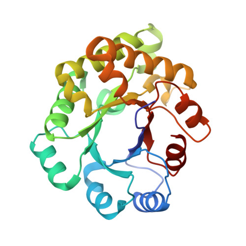
Loop 8 (residues 232-242) in triosephosphate isomerase (TIM) is a highly conserved loop that forms a tight binding pocket for the phosphate moiety of the substrate. Its sequence includes the fully conserved, solvent-exposed Leu238. The tight phosphate-binding pocket explains the high substrate specificity of TIM being limited to the in vivo substrates dihydroxyacetone-phosphate and D-glyceraldehyde-3-phosphate. Here we use the monomeric variant of trypanosomal TIM for exploring the structural consequences of shortening this loop. The mutagenesis, guided by extensive modeling calculations and followed up by crystallographic characterization, is aimed at widening the phosphate-binding pocket and, consequently, changing the substrate specificity. Two new variants were characterized. The crystal structures of these variants indicate that in monomeric forms of TIM, the Leu238 side-chain is nicely buried in a hydrophobic cluster. Monomeric forms of wild-type dimeric TIM are known to exist transiently as folding intermediates; our structural analysis suggests that in this monomeric form, Leu238 of loop 8 also adopts this completely buried conformation, which explains its full conservation across the evolution. The much wider phosphate-binding pocket of the new variant allows for the development of a new TIM variant with a different substrate specificity.
Organizational Affiliation:
Biocenter Oulu and Department of Biochemistry, University of Oulu, Linnanmaa, Oulu, Finland.