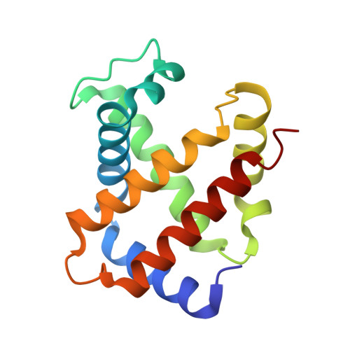
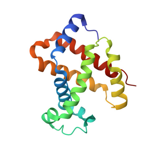
Oxygen binding by alpha(Fe2+)2beta(Ni2+)2 hemoglobin crystals.
Bruno, S., Bettati, S., Manfredini, M., Mozzarelli, A., Bolognesi, M., Deriu, D., Rosano, C., Tsuneshige, A., Yonetani, T., Henry, E.R.(2000) Protein Sci 9: 683-692
- PubMed: 10794410
- DOI: https://doi.org/10.1110/ps.9.4.683
- Primary Citation of Related Structures:
1DKE - PubMed Abstract:
Oxygen binding by hemoglobin fixed in the T state either by crystallization or by encapsulation in silica gels is apparently noncooperative. However, cooperativity might be masked by different oxygen affinities of alpha and beta subunits. Metal hybrid hemoglobins, where the noniron metal does not bind oxygen, provide the opportunity to determine the oxygen affinities of alpha and beta hemes separately. Previous studies have characterized the oxygen binding by alpha(Ni2+)2beta(Fe2+)2 crystals. Here, we have determined the three-dimensional (3D) structure and oxygen binding of alpha(Fe2+)2beta(Ni2+)2 crystals grown from polyethylene glycol solutions. Polarized absorption spectra were recorded at different oxygen pressures with light polarized parallel either to the b or c crystal axis by single crystal microspectrophotometry. The oxygen pressures at 50% saturation (p50s) are 95 +/- 3 and 87 +/- 4 Torr along the b and c crystal axes, respectively, and the corresponding Hill coefficients are 0.96 +/- 0.06 and 0.90 +/- 0.03. Analysis of the binding curves, taking into account the different projections of the alpha hemes along the optical directions, indicates that the oxygen affinity of alpha1 hemes is 1.3-fold lower than alpha2 hemes. Inspection of the 3D structure suggests that this inequivalence may arise from packing interactions of the Hb tetramer within the monoclinic crystal lattice. A similar inequivalence was found for the beta subunits of alpha(Ni2+)2beta(Fe2+)2 crystals. The average oxygen affinity of the alpha subunits (p50 = 91 Torr) is about 1.2-fold higher than the beta subunits (p50 = 110 Torr). In the absence of cooperativity, this heterogeneity yields an oxygen binding curve of Hb A with a Hill coefficient of 0.999. Since the binding curves of Hb A crystals exhibit a Hill coefficient very close to unity, these findings indicate that oxygen binding by T-state hemoglobin is noncooperative, in keeping with the Monod, Wyman, and Changeux model.
Organizational Affiliation:
Institute of Biochemical Sciences, University of Parma, Italy.