X-ray structure of beta-carbonic anhydrase from the red alga, Porphyridium purpureum, reveals a novel catalytic site for CO(2) hydration.
Mitsuhashi, S., Mizushima, T., Yamashita, E., Yamamoto, M., Kumasaka, T., Moriyama, H., Ueki, T., Miyachi, S., Tsukihara, T.(2000) J Biol Chem 275: 5521-5526
- PubMed: 10681531
- DOI: https://doi.org/10.1074/jbc.275.8.5521
- Primary Citation of Related Structures:
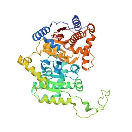
1DDZ - PubMed Abstract:
The carbonic anhydrases (CAs) fall into three evolutionarily distinct families designated alpha-, beta-, and gamma-CAs based on their primary structure. beta-CAs are present in higher plants, algae, and prokaryotes, and are involved in inorganic carbon utilization. Here, we describe the novel x-ray structure of beta-CA from the red alga, Porphyridium purpureum, at 2.2-A resolution using intrinsic zinc multiwavelength anomalous diffraction phasing. The CA monomer is composed of two internally repeating structures, being folded as a pair of fundamentally equivalent motifs of an alpha/beta domain and three projecting alpha-helices. The motif is obviously distinct from that of either alpha- or gamma-CAs. This homodimeric CA appears like a tetramer with a pseudo 222 symmetry. The active site zinc is coordinated by a Cys-Asp-His-Cys tetrad that is strictly conserved among the beta-CAs. No water molecule is found in a zinc-liganding radius, indicating that the zinc-hydroxide mechanism in alpha-CAs, and possibly in gamma-CAs, is not directly applicable to the case in beta-CAs. Zinc coordination environments of the CAs provide an interesting example of the convergent evolution of distinct catalytic sites required for the same CO(2) hydration reaction.
Organizational Affiliation:
Marine Biotechnology Institute, Kamaishi Laboratories, Heita, Kamaishi, Iwate, 026-0001 Japan. mitsuhashi@kamaishi.mbio.co.jp