1DAA

CRYSTALLOGRAPHIC STRUCTURE OF D-AMINO ACID AMINOTRANSFERASE COMPLEXED WITH PYRIDOXAL-5'-PHOSPHATE


Experimental Data Snapshot

  • Method: X-RAY DIFFRACTION
  • Resolution: 1.94 Å
  • R-Value Work: 0.184 
  • R-Value Observed: 0.184 

wwPDB Validation   3D Report Full Report


This is version 1.4 of the entry. See complete history


Literature

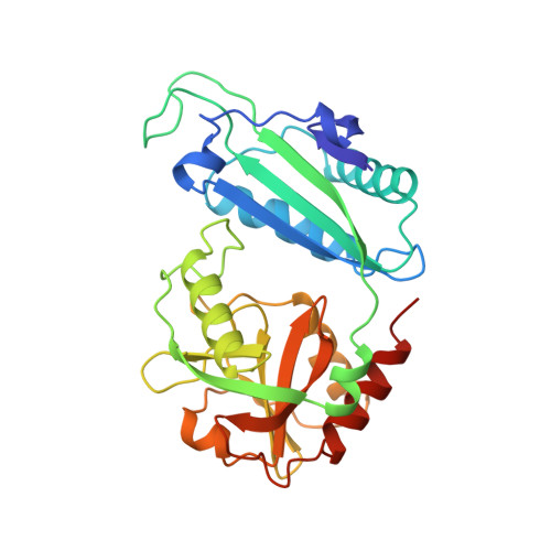
Crystal structure of a D-amino acid aminotransferase: how the protein controls stereoselectivity.

Sugio, S.Petsko, G.A.Manning, J.M.Soda, K.Ringe, D.

(1995) Biochemistry 34: 9661-9669

  • DOI: https://doi.org/10.1021/bi00030a002
  • Primary Citation of Related Structures:  
    1DAA

  • PubMed Abstract: 

    The three-dimensional structure of D-amino acid aminotransferase (D-AAT) in the pyridoxamine phosphate form has been determined crystallographically. The fold of this pyridoxal phosphate (PLP)-containing enzyme is completely different from those of any of the other enzymes that utilize PLP as part of their mechanism and whose structures are known. However, there are some striking similarities between the active sites of D-AAT and the corresponding enzyme that transaminates L-amino acids, L-aspartate aminotransferase. These similarities represent convergent evolution to a common solution of the problem of enforcing transamination chemistry on the PLP cofactor. Implications of these similarities are discussed in terms of their possible roles in the stabilization of intermediates of a transamination reaction. In addition, sequence similarity between D-AAT and branched chain L-amino acid aminotransferase suggests that this latter enzyme will also have a fold similar to that of D-AAT.


  • Organizational Affiliation

    Department of Biochemistry, Brandeis University, Waltham, Massachusetts 02254, USA.


Macromolecules
Find similar proteins by:  (by identity cutoff)  |  3D Structure
Entity ID: 1
MoleculeChains Sequence LengthOrganismDetailsImage
D-AMINO ACID AMINOTRANSFERASE
A, B
282Bacillus sp. YM-1Mutation(s): 0 
EC: 2.6.1.21
UniProt
Find proteins for P19938 (Bacillus sp. (strain YM-1))
Explore P19938 
Go to UniProtKB:  P19938
Entity Groups  
Sequence Clusters30% Identity50% Identity70% Identity90% Identity95% Identity100% Identity
UniProt GroupP19938
Sequence Annotations
Expand
  • Reference Sequence
Small Molecules
Ligands 1 Unique
IDChains Name / Formula / InChI Key2D Diagram3D Interactions
PLP
Query on PLP

Download Ideal Coordinates CCD File 
C [auth A],
D [auth B]
PYRIDOXAL-5'-PHOSPHATE
C8 H10 N O6 P
NGVDGCNFYWLIFO-UHFFFAOYSA-N
Experimental Data & Validation

Experimental Data

  • Method: X-RAY DIFFRACTION
  • Resolution: 1.94 Å
  • R-Value Work: 0.184 
  • R-Value Observed: 0.184 
  • Space Group: P 1 21 1
Unit Cell:
Length ( Å )Angle ( ˚ )
a = 58.5α = 90
b = 76.2β = 103.9
c = 73.3γ = 90
Software Package:
Software NamePurpose
X-PLORmodel building
X-PLORrefinement
X-PLORphasing

Structure Validation

View Full Validation Report


Entry History 

Deposition Data

Revision History  (Full details and data files)

  • Version 1.0: 1995-09-15
    Type: Initial release
  • Version 1.1: 2008-03-03
    Changes: Version format compliance
  • Version 1.2: 2011-07-13
    Changes: Source and taxonomy, Version format compliance
  • Version 1.3: 2018-04-04
    Changes: Advisory, Data collection, Other
  • Version 1.4: 2024-02-07
    Changes: Advisory, Data collection, Database references, Derived calculations