Crystal structure of Escherichia coli malate synthase G complexed with magnesium and glyoxylate at 2.0 A resolution: mechanistic implications.
Howard, B.R., Endrizzi, J.A., Remington, S.J.(2000) Biochemistry 39: 3156-3168
- PubMed: 10715138
- DOI: https://doi.org/10.1021/bi992519h
- Primary Citation of Related Structures:
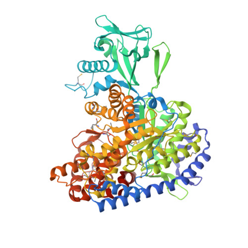
1D8C - PubMed Abstract:
The crystal structure of selenomethionine-substituted malate synthase G, an 81 kDa monomeric enzyme from Escherichia coli has been determined by MAD phasing, model building, and crystallographic refinement to a resolution of 2.0 A. The crystallographic R factor is 0.177 for 49 242 reflections observed at the incident wavelength of 1.008 A, and the model stereochemistry is satisfactory. The basic fold of the enzyme is that of a beta8/alpha8 (TIM) barrel. The barrel is centrally located, with an N-terminal alpha-helical domain flanking one side. An inserted beta-sheet domain folds against the opposite side of the barrel, and an alpha-helical C-terminal domain forms a plug which caps the active site. Malate synthase catalyzes the condensation of glyoxylate and acetyl-coenzyme A and hydrolysis of the intermediate to yield malate and coenzyme A, requiring Mg(2+). The structure reveals an enzyme-substrate complex with glyoxylate and Mg(2+) which coordinates the aldehyde and carboxylate functions of the substrate. Two strictly conserved residues, Asp631 and Arg338, are proposed to provide concerted acid-base chemistry for the generation of the enol(ate) intermediate of acetyl-coenzyme A, while main-chain hydrogen bonds and bound Mg(2+) polarize glyoxylate in preparation for nucleophilic attack. The catalytic strategy of malate synthase appears to be essentially the same as that of citrate synthase, with the electrophile activated for nucleophilic attack by nearby positive charges and hydrogen bonds, while concerted acid-base catalysis accomplishes the abstraction of a proton from the methyl group of acetyl-coenzyme A. An active site aspartate is, however, the only common feature of these two enzymes, and the active sites of these enzymes are produced by quite different protein folds. Interesting similarities in the overall folds and modes of substrate recognition are discussed in comparisons of malate synthase with pyruvate kinase and pyruvate phosphate dikinase.
Organizational Affiliation:
Institute of Molecular Biology and Departments of Chemistry and Physics, University of Oregon, Eugene, Oregon 97403, USA.