1CZF

ENDO-POLYGALACTURONASE II FROM ASPERGILLUS NIGER


Experimental Data Snapshot

  • Method: X-RAY DIFFRACTION
  • Resolution: 1.68 Å
  • R-Value Free: 0.191 
  • R-Value Work: 0.166 
  • R-Value Observed: 0.166 

wwPDB Validation   3D Report Full Report


Ligand Structure Quality Assessment 


This is version 1.3 of the entry. See complete history


Literature

1.68-A crystal structure of endopolygalacturonase II from Aspergillus niger and identification of active site residues by site-directed mutagenesis.

van Santen, Y.Benen, J.A.Schroter, K.H.Kalk, K.H.Armand, S.Visser, J.Dijkstra, B.W.

(1999) J Biol Chem 274: 30474-30480

  • DOI: https://doi.org/10.1074/jbc.274.43.30474
  • Primary Citation of Related Structures:  
    1CZF

  • PubMed Abstract: 

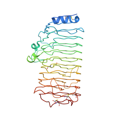
    Polygalacturonases specifically hydrolyze polygalacturonate, a major constituent of plant cell wall pectin. To understand the catalytic mechanism and substrate and product specificity of these enzymes, we have solved the x-ray structure of endopolygalacturonase II of Aspergillus niger and we have carried out site-directed mutagenesis studies. The enzyme folds into a right-handed parallel beta-helix with 10 complete turns. The beta-helix is composed of four parallel beta-sheets, and has one very small alpha-helix near the N terminus, which shields the enzyme's hydrophobic core. Loop regions form a cleft on the exterior of the beta-helix. Site-directed mutagenesis of Asp(180), Asp(201), Asp(202), His(223), Arg(256), and Lys(258), which are located in this cleft, results in a severe reduction of activity, demonstrating that these residues are important for substrate binding and/or catalysis. The juxtaposition of the catalytic residues differs from that normally encountered in inverting glycosyl hydrolases. A comparison of the endopolygalacturonase II active site with that of the P22 tailspike rhamnosidase suggests that Asp(180) and Asp(202) activate the attacking nucleophilic water molecule, while Asp(201) protonates the glycosidic oxygen of the scissile bond.


  • Organizational Affiliation

    Laboratory of Biophysical Chemistry, Groningen University, 9747 AG Groningen, The Netherlands.


Macromolecules
Find similar proteins by:  (by identity cutoff)  |  3D Structure
Entity ID: 1
MoleculeChains Sequence LengthOrganismDetailsImage
POLYGALACTURONASE II
A, B
362Aspergillus nigerMutation(s): 0 
EC: 3.2.1.15
UniProt
Find proteins for P26214 (Aspergillus niger (strain ATCC 1015 / CBS 113.46 / FGSC A1144 / LSHB Ac4 / NCTC 3858a / NRRL 328 / USDA 3528.7))
Explore P26214 
Go to UniProtKB:  P26214
Entity Groups  
Sequence Clusters30% Identity50% Identity70% Identity90% Identity95% Identity100% Identity
UniProt GroupP26214
Sequence Annotations
Expand
  • Reference Sequence
Experimental Data & Validation

Experimental Data

  • Method: X-RAY DIFFRACTION
  • Resolution: 1.68 Å
  • R-Value Free: 0.191 
  • R-Value Work: 0.166 
  • R-Value Observed: 0.166 
  • Space Group: P 21 21 2
Unit Cell:
Length ( Å )Angle ( ˚ )
a = 65.497α = 90
b = 201.241β = 90
c = 49.065γ = 90
Software Package:
Software NamePurpose
DENZOdata reduction
SCALEPACKdata scaling
PHASESphasing
WARPmodel building
X-PLORrefinement
ARP/wARPmodel building

Structure Validation

View Full Validation Report


Ligand Structure Quality Assessment 


Entry History 

Deposition Data

Revision History  (Full details and data files)

  • Version 1.0: 1999-10-28
    Type: Initial release
  • Version 1.1: 2007-10-16
    Changes: Version format compliance
  • Version 1.2: 2011-07-13
    Changes: Derived calculations, Version format compliance
  • Version 1.3: 2020-07-29
    Type: Remediation
    Reason: Carbohydrate remediation
    Changes: Data collection, Derived calculations, Structure summary