1CSK

THE CRYSTAL STRUCTURE OF HUMAN CSKSH3: STRUCTURAL DIVERSITY NEAR THE RT-SRC AND N-SRC LOOP


Experimental Data Snapshot

  • Method: X-RAY DIFFRACTION
  • Resolution: 2.50 Å
  • R-Value Free: 0.278 
  • R-Value Work: 0.224 
  • R-Value Observed: 0.224 

wwPDB Validation   3D Report Full Report


This is version 1.3 of the entry. See complete history


Literature

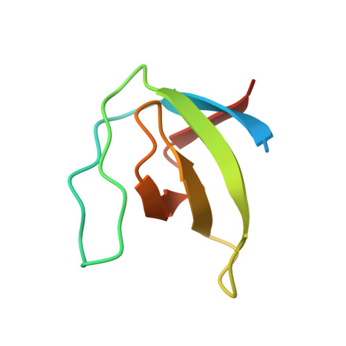
The crystal structure of human CskSH3: structural diversity near the RT-Src and n-Src loop.

Borchert, T.V.Mathieu, M.Zeelen, J.P.Courtneidge, S.A.Wierenga, R.K.

(1994) FEBS Lett 341: 79-85

  • DOI: https://doi.org/10.1016/0014-5793(94)80244-0
  • Primary Citation of Related Structures:  
    1CSK

  • PubMed Abstract: 

    SH3 domains are modules occurring in diverse proteins, ranging from cytoskeletal proteins to signaling proteins, such as tyrosine kinases. The crystal structure of the SH3 domain of Csk (c-Src specific tyrosine kinase) has been refined at a resolution of 2.5 A, with an R-factor of 22.4%. The structure is very similar to the FynSH3 crystal structure. When comparing CskSH3 and FynSH3 it is seen that the structural and charge differences of the RT-Src loop and the n-Src loop, near the conserved Trp47, correlate with different binding properties of these SH3 domains. The structure comparison suggests that those glycines and acid residues which are very well conserved in the SH3 sequences are important for the stability of the SH3 fold.


  • Organizational Affiliation

    EMBL, Heidelberg, Germany.


Macromolecules
Find similar proteins by:  (by identity cutoff)  |  3D Structure
Entity ID: 1
MoleculeChains Sequence LengthOrganismDetailsImage
C-SRC SH3 DOMAIN
A, B, C, D
71Homo sapiensMutation(s): 0 
EC: 2.7.1.112
UniProt & NIH Common Fund Data Resources
Find proteins for P41240 (Homo sapiens)
Explore P41240 
Go to UniProtKB:  P41240
PHAROS:  P41240
GTEx:  ENSG00000103653 
Entity Groups  
Sequence Clusters30% Identity50% Identity70% Identity90% Identity95% Identity100% Identity
UniProt GroupP41240
Sequence Annotations
Expand
  • Reference Sequence
Experimental Data & Validation

Experimental Data

  • Method: X-RAY DIFFRACTION
  • Resolution: 2.50 Å
  • R-Value Free: 0.278 
  • R-Value Work: 0.224 
  • R-Value Observed: 0.224 
  • Space Group: C 2 2 21
Unit Cell:
Length ( Å )Angle ( ˚ )
a = 64.9α = 90
b = 74.69β = 90
c = 125.03γ = 90
Software Package:
Software NamePurpose
X-PLORmodel building
X-PLORrefinement
X-PLORphasing

Structure Validation

View Full Validation Report


Entry History 

Deposition Data

Revision History  (Full details and data files)

  • Version 1.0: 1994-07-31
    Type: Initial release
  • Version 1.1: 2008-03-24
    Changes: Version format compliance
  • Version 1.2: 2011-07-13
    Changes: Version format compliance
  • Version 1.3: 2024-02-07
    Changes: Data collection, Database references, Other