1CSC

Structure of ternary complexes of citrate synthase with D-and L-malate: Mechanistic implications


Experimental Data Snapshot

  • Method: X-RAY DIFFRACTION
  • Resolution: 1.70 Å
  • R-Value Observed: 0.188 

wwPDB Validation   3D Report Full Report


This is version 1.4 of the entry. See complete history


Literature

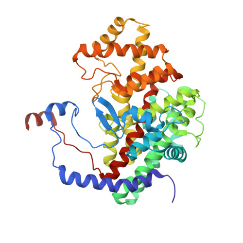
1.9-A structures of ternary complexes of citrate synthase with D- and L-malate: mechanistic implications.

Karpusas, M.Holland, D.Remington, S.J.

(1991) Biochemistry 30: 6024-6031

  • DOI: https://doi.org/10.1021/bi00238a028
  • Primary Citation of Related Structures:  
    1CSC, 2CSC, 3CSC, 4CSC

  • PubMed Abstract: 

    The structures of four isomorphous crystals of ternary complexes of chicken heart citrate synthase with D- or L-malate and acetyl coenzyme A or carboxymethyl coenzyme A have been determined by X-ray crystallography and fully refined at 1.9-A resolution. The structures show that both L-malate and D-malate bind in a very similar way in the presence of acetylCoA and that the enzyme conformation is "closed". Hydrogen bond geometry is suggested to account for the difference in binding constants of the two stereoisomers. The structures suggest that steric hindrance can account for the observation that proton exchange of acetyl coenzyme A with solvent is catalyzed by citrate synthase in the presence of L-malate but not D-malate. The ternary complexes with malate reveal the mode of binding of the substrate acetylCoA in the ground state. The carbonyl oxygen of the acetyl group is hydrogen bonded to a water molecule and to histidine 274, allowing unambiguous identification of the orientation of this group. The structures support the hypothesis that carboxymethyl coenzyme A is a transition-state analogue for the enolization step of the reaction (Bayer et al., 1981) and additionally support proposed mechanisms for the condensation reaction (Karpusas et al., 1990; Alter et al., 1990).


  • Organizational Affiliation

    Institute of Molecular Biology, University of Oregon, Eugene 97403.


Macromolecules
Find similar proteins by:  (by identity cutoff)  |  3D Structure
Entity ID: 1
MoleculeChains Sequence LengthOrganismDetailsImage
CITRATE SYNTHASE433Gallus gallusMutation(s): 0 
EC: 4.1.3.7
UniProt
Find proteins for P23007 (Gallus gallus)
Explore P23007 
Go to UniProtKB:  P23007
Entity Groups  
Sequence Clusters30% Identity50% Identity70% Identity90% Identity95% Identity100% Identity
UniProt GroupP23007
Sequence Annotations
Expand
  • Reference Sequence
Small Molecules
Ligands 2 Unique
IDChains Name / Formula / InChI Key2D Diagram3D Interactions
CMC
Query on CMC

Download Ideal Coordinates CCD File 
B [auth A]CARBOXYMETHYL COENZYME *A
C23 H38 N7 O18 P3 S
OBUOSIHPWVNVJN-GRFIIANRSA-N
LMR
Query on LMR

Download Ideal Coordinates CCD File 
C [auth A](2S)-2-hydroxybutanedioic acid
C4 H6 O5
BJEPYKJPYRNKOW-REOHCLBHSA-N
Experimental Data & Validation

Experimental Data

  • Method: X-RAY DIFFRACTION
  • Resolution: 1.70 Å
  • R-Value Observed: 0.188 
  • Space Group: C 1 2 1
Unit Cell:
Length ( Å )Angle ( ˚ )
a = 104α = 90
b = 78.1β = 78.9
c = 58.3γ = 90
Software Package:
Software NamePurpose
TNTrefinement

Structure Validation

View Full Validation Report


Entry History 

Deposition Data

Revision History  (Full details and data files)

  • Version 1.0: 1991-04-15
    Type: Initial release
  • Version 1.1: 2008-03-24
    Changes: Version format compliance
  • Version 1.2: 2011-07-13
    Changes: Version format compliance
  • Version 1.3: 2017-11-29
    Changes: Derived calculations, Other
  • Version 1.4: 2024-02-07
    Changes: Data collection, Database references, Derived calculations