1CPS

STRUCTURAL COMPARISON OF SULFODIIMINE AND SULFONAMIDE INHIBITORS IN THEIR COMPLEXES WITH ZINC ENZYMES


Experimental Data Snapshot

  • Method: X-RAY DIFFRACTION
  • Resolution: 2.25 Å
  • R-Value Observed: 0.174 

wwPDB Validation   3D Report Full Report


This is version 1.2 of the entry. See complete history


Literature

Structural comparison of sulfodiimine and sulfonamide inhibitors in their complexes with zinc enzymes.

Cappalonga, A.M.Alexander, R.S.Christianson, D.W.

(1992) J Biol Chem 267: 19192-19197

  • DOI: https://doi.org/10.2210/pdb1cps/pdb
  • Primary Citation of Related Structures:  
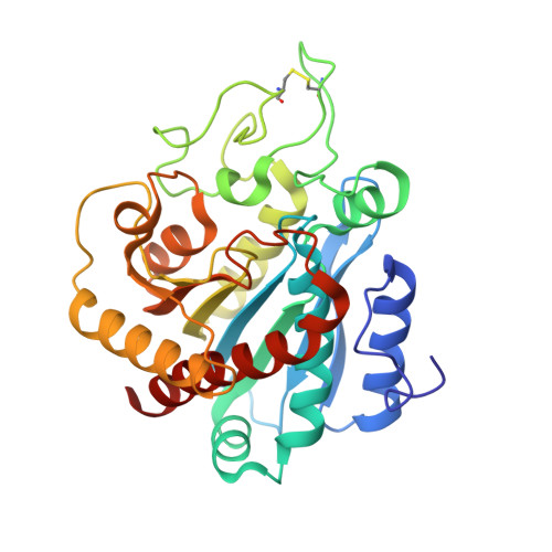
    1CPS

  • PubMed Abstract: 

    The three-dimensional structure of (L(-)-2-carboxy-3-phenylpropyl) methylsulfodiimine in its complex with the zinc metalloenzyme carboxypeptidase A has been determined at 2.25-A resolution by x-ray crystallographic methods. This is the first example of a sulfodiimine-containing inhibitor binding to a zinc enzyme, and the structure of the enzyme-inhibitor complex reveals that the tetrahedral sulfodiimine group coordinates to the active site zinc ion in unidentate fashion. The zinc-coordinated nitrogen atom of the sulfodiimine group is also within hydrogen bonding distance to active site base Glu-270; presumably, the sulfodiimine is ionized and accepts a hydrogen bond from protonated Glu-270. The other sulfodiimine nitrogen accepts a hydrogen bond from Arg-127, and the inhibitor binds as a possible analogue of the tetrahedral transition state (or intermediate) in a promoted water pathway for peptide hydrolysis. The unidentate sulfodiimine-zinc binding mode observed in this enzyme-inhibitor complex is reminiscent of that observed in sulfonamide complexes with the zinc metalloenzyme carbonic anhydrase II, and the structural features of sulfodiimine- and sulfonamide-zinc interactions exhibit important similarities among recently determined structures of enzyme-inhibitor complexes: ionized nitrogens bind to zinc in each structure, and these nitrogens are engaged in hydrogen bond interactions with neighboring enzyme residues.


  • Organizational Affiliation

    Department of Chemistry, University of Pennsylvania, Philadelphia 19104-6323.


Macromolecules
Find similar proteins by:  (by identity cutoff)  |  3D Structure
Entity ID: 1
MoleculeChains Sequence LengthOrganismDetailsImage
CARBOXYPEPTIDASE A307Bos taurusMutation(s): 0 
EC: 3.4.17.1
UniProt
Find proteins for P00730 (Bos taurus)
Explore P00730 
Go to UniProtKB:  P00730
Entity Groups  
Sequence Clusters30% Identity50% Identity70% Identity90% Identity95% Identity100% Identity
UniProt GroupP00730
Sequence Annotations
Expand
  • Reference Sequence
Small Molecules
Ligands 2 Unique
IDChains Name / Formula / InChI Key2D Diagram3D Interactions
CPM
Query on CPM

Download Ideal Coordinates CCD File 
C [auth A]S-(2-CARBOXY-3-PHENYLPROPYL)THIODIIMINE-S-METHANE
C11 H16 N2 O2 S
FKHYVJWYNYZPCA-SNVBAGLBSA-N
ZN
Query on ZN

Download Ideal Coordinates CCD File 
B [auth A]ZINC ION
Zn
PTFCDOFLOPIGGS-UHFFFAOYSA-N
Binding Affinity Annotations 
IDSourceBinding Affinity
CPM Binding MOAD:  1CPS Ki: 220 (nM) from 1 assay(s)
PDBBind:  1CPS Ki: 220 (nM) from 1 assay(s)
Experimental Data & Validation

Experimental Data

  • Method: X-RAY DIFFRACTION
  • Resolution: 2.25 Å
  • R-Value Observed: 0.174 
  • Space Group: P 1 21 1
Unit Cell:
Length ( Å )Angle ( ˚ )
a = 51.7α = 90
b = 60.4β = 97.4
c = 47.2γ = 90
Software Package:
Software NamePurpose
PROLSQrefinement

Structure Validation

View Full Validation Report


Entry History 

Deposition Data

Revision History  (Full details and data files)

  • Version 1.0: 1993-10-31
    Type: Initial release
  • Version 1.1: 2008-03-24
    Changes: Version format compliance
  • Version 1.2: 2011-07-13
    Changes: Version format compliance