1CN3

INTERACTION OF POLYOMAVIRUS INTERNAL PROTEIN VP2 WITH MAJOR CAPSID PROTEIN VP1 AND IMPLICATIONS FOR PARTICIPATION OF VP2 IN VIRAL ENTRY


Experimental Data Snapshot

  • Method: X-RAY DIFFRACTION
  • Resolution: 2.20 Å
  • R-Value Free: 0.260 
  • R-Value Work: 0.230 

wwPDB Validation   3D Report Full Report


This is version 1.3 of the entry. See complete history


Literature

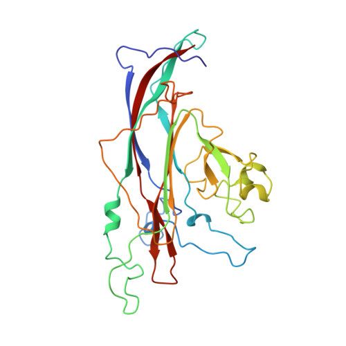
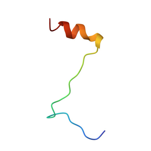
Interaction of polyomavirus internal protein VP2 with the major capsid protein VP1 and implications for participation of VP2 in viral entry.

Chen, X.S.Stehle, T.Harrison, S.C.

(1998) EMBO J 17: 3233-3240

  • DOI: https://doi.org/10.1093/emboj/17.12.3233
  • Primary Citation of Related Structures:  
    1CN3

  • PubMed Abstract: 

    A complex of the polyomavirus internal protein VP2/VP3 with the pentameric major capsid protein VP1 has been prepared by co-expression in Escherichia coli. A C-terminal segment of VP2/VP3 is required for tight association, and a crystal structure of this segment, complexed with a VP1 pentamer, has been determined at 2.2 A resolution. The structure shows specific contacts between a single copy of the internal protein and a pentamer of VP1. These interactions were not detected in the previously described structure of the virion, but the location of VP2 in the recombinant complex is consistent with features in the virion electron-density map. The C-terminus of VP2/VP3 inserts in an unusual, hairpin-like manner into the axial cavity of the VP1 pentamer, where it is anchored strongly by hydrophobic interactions. The remainder of the internal protein appears to have significant flexibility. This structure restricts possible models for exposure of the internal proteins during viral entry.


  • Organizational Affiliation

    Howard Hughes Medical Institute and Department of Molecular and Cellular Biology, Harvard University, Boston, MA 02138, USA.


Macromolecules
Find similar proteins by:  (by identity cutoff)  |  3D Structure
Entity ID: 1
MoleculeChains Sequence LengthOrganismDetailsImage
COAT PROTEIN VP1
A, B, C, D, E
283Polyomavirus sp.Mutation(s): 0 
UniProt
Find proteins for P49302 (Murine polyomavirus (strain P16 small-plaque))
Explore P49302 
Go to UniProtKB:  P49302
Entity Groups  
Sequence Clusters30% Identity50% Identity70% Identity90% Identity95% Identity100% Identity
UniProt GroupP49302
Sequence Annotations
Expand
  • Reference Sequence
Find similar proteins by:  (by identity cutoff)  |  3D Structure
Entity ID: 2
MoleculeChains Sequence LengthOrganismDetailsImage
FRAGMENT OF COAT PROTEIN VP229N/AMutation(s): 0 
UniProt
Find proteins for P12908 (Murine polyomavirus (strain Crawford small-plaque))
Explore P12908 
Go to UniProtKB:  P12908
Entity Groups  
Sequence Clusters30% Identity50% Identity70% Identity90% Identity95% Identity100% Identity
UniProt GroupP12908
Sequence Annotations
Expand
  • Reference Sequence
Experimental Data & Validation

Experimental Data

  • Method: X-RAY DIFFRACTION
  • Resolution: 2.20 Å
  • R-Value Free: 0.260 
  • R-Value Work: 0.230 
  • Space Group: P 31 2 1
Unit Cell:
Length ( Å )Angle ( ˚ )
a = 219α = 90
b = 219β = 90
c = 99γ = 120
Software Package:
Software NamePurpose
X-PLORrefinement

Structure Validation

View Full Validation Report


Entry History 

Deposition Data

Revision History  (Full details and data files)

  • Version 1.0: 1999-06-09
    Type: Initial release
  • Version 1.1: 2008-04-26
    Changes: Version format compliance
  • Version 1.2: 2011-07-13
    Changes: Version format compliance
  • Version 1.3: 2023-12-27
    Changes: Data collection, Database references