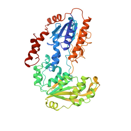
1CJC

STRUCTURE OF ADRENODOXIN REDUCTASE OF MITOCHONDRIAL P450 SYSTEMS


Experimental Data Snapshot

  • Method: X-RAY DIFFRACTION
  • Resolution: 1.70 Å
  • R-Value Free: 0.223 
  • R-Value Work: 0.188 

wwPDB Validation   3D Report Full Report


Ligand Structure Quality Assessment 


This is version 1.5 of the entry. See complete history


Literature

The structure of adrenodoxin reductase of mitochondrial P450 systems: electron transfer for steroid biosynthesis.

Ziegler, G.A.Vonrhein, C.Hanukoglu, I.Schulz, G.E.

(1999) J Mol Biol 289: 981-990

  • DOI: https://doi.org/10.1006/jmbi.1999.2807
  • Primary Citation of Related Structures:  
    1CJC

  • PubMed Abstract: 

    Adrenodoxin reductase is a monomeric 51 kDa flavoenzyme that is involved in the biosynthesis of all steroid hormones. The structure of the native bovine enzyme was determined at 2.8 A resolution, and the structure of the respective recombinant enzyme at 1.7 A resolution. Adrenodoxin reductase receives a two-electron package from NADPH and converts it to two single electrons that are transferred via adrenodoxin to all mitochondrial cytochromes P 450. The structure suggests how the observed flavin semiquinone is stabilized. A striking feature is the asymmetric charge distribution, which most likely controls the approach of the electron carrier adrenodoxin. A model for the interaction is proposed. Adrenodoxin reductase shows clear sequence homology to half a dozen proteins identified in genome analysis projects, but neither sequence nor structural homology to established, functionally related electron transferases. Yet, the structure revealed a relationship to the disulfide oxidoreductases, permitting the assignment of the NADP-binding site.


  • Organizational Affiliation

    Albert-Ludwigs-Universität, Albertstrasse 21, Freiburg im Breisgau, D-79104, Germany.


Macromolecules
Find similar proteins by:  (by identity cutoff)  |  3D Structure
Entity ID: 1
MoleculeChains Sequence LengthOrganismDetailsImage
PROTEIN (ADRENODOXIN REDUCTASE)460Bos taurusMutation(s): 0 
EC: 1.18.1.6
UniProt
Find proteins for P08165 (Bos taurus)
Explore P08165 
Go to UniProtKB:  P08165
Entity Groups  
Sequence Clusters30% Identity50% Identity70% Identity90% Identity95% Identity100% Identity
UniProt GroupP08165
Sequence Annotations
Expand
  • Reference Sequence
Small Molecules
Ligands 1 Unique
IDChains Name / Formula / InChI Key2D Diagram3D Interactions
FAD
Query on FAD

Download Ideal Coordinates CCD File 
B [auth A]FLAVIN-ADENINE DINUCLEOTIDE
C27 H33 N9 O15 P2
VWWQXMAJTJZDQX-UYBVJOGSSA-N
Experimental Data & Validation

Experimental Data

  • Method: X-RAY DIFFRACTION
  • Resolution: 1.70 Å
  • R-Value Free: 0.223 
  • R-Value Work: 0.188 
  • Space Group: P 1 21 1
Unit Cell:
Length ( Å )Angle ( ˚ )
a = 60.81α = 90
b = 62.53β = 106.76
c = 78.37γ = 90
Software Package:
Software NamePurpose
SHARPphasing
REFMACrefinement
MOSFLMdata reduction
CCP4data scaling

Structure Validation

View Full Validation Report


Ligand Structure Quality Assessment 


Entry History 

Deposition Data

Revision History  (Full details and data files)

  • Version 1.0: 1999-04-30
    Type: Initial release
  • Version 1.1: 2008-04-26
    Changes: Version format compliance
  • Version 1.2: 2011-07-13
    Changes: Version format compliance
  • Version 1.3: 2013-04-17
    Changes: Structure summary
  • Version 1.4: 2019-11-27
    Changes: Advisory, Data collection, Database references
  • Version 1.5: 2023-12-27
    Changes: Advisory, Data collection, Database references, Derived calculations