1CIP

GI-ALPHA-1 SUBUNIT OF GUANINE NUCLEOTIDE-BINDING PROTEIN COMPLEXED WITH A GTP ANALOGUE


Experimental Data Snapshot

  • Method: X-RAY DIFFRACTION
  • Resolution: 1.50 Å
  • R-Value Free: 0.235 
  • R-Value Work: 0.213 

wwPDB Validation   3D Report Full Report


Ligand Structure Quality Assessment 


This is version 1.3 of the entry. See complete history


Literature

Structure of Gialpha1.GppNHp, autoinhibition in a galpha protein-substrate complex.

Coleman, D.E.Sprang, S.R.

(1999) J Biol Chem 274: 16669-16672

  • DOI: https://doi.org/10.1074/jbc.274.24.16669
  • Primary Citation of Related Structures:  
    1CIP

  • PubMed Abstract: 

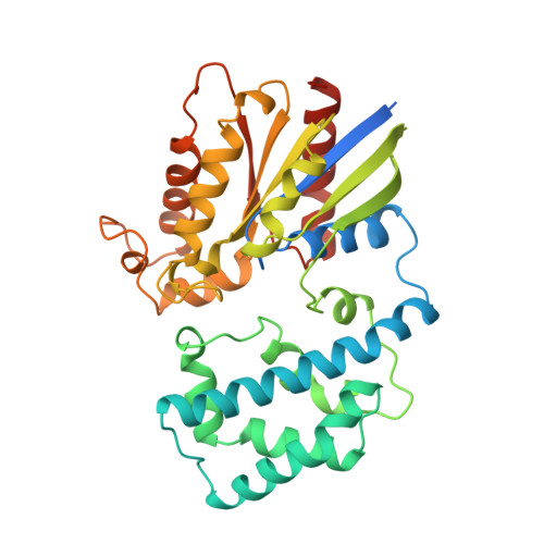
    The structure of the G protein Gialpha1 complexed with the nonhydrolyzable GTP analog guanosine-5'-(betagamma-imino)triphosphate (GppNHp) has been determined at a resolution of 1.5 A. In the active site of Gialpha1. GppNHp, a water molecule is hydrogen bonded to the side chain of Glu43 and to an oxygen atom of the gamma-phosphate group. The side chain of the essential catalytic residue Gln204 assumes a conformation which is distinctly different from that observed in complexes with either guanosine 5'-O-3-thiotriphosphate or the transition state analog GDP.AlF4-. Hydrogen bonding and steric interactions position Gln204 such that it interacts with a presumptive nucleophilic water molecule, but cannot interact with the pentacoordinate transition state. Gln204 must be released from this auto-inhibited state to participate in catalysis. RGS proteins may accelerate the rate of GTP hydrolysis by G protein alpha subunits, in part, by inserting an amino acid side chain into the site occupied by Gln204, thereby destabilizing the auto-inhibited state of Galpha.


  • Organizational Affiliation

    Department of Biochemistry, The University of Texas Southwestern Medical Center, Dallas, Texas 75235-9050, USA.


Macromolecules
Find similar proteins by:  (by identity cutoff)  |  3D Structure
Entity ID: 1
MoleculeChains Sequence LengthOrganismDetailsImage
PROTEIN (GUANINE NUCLEOTIDE-BINDING PROTEIN ALPHA-1 SUBUNIT)353Rattus norvegicusMutation(s): 0 
UniProt
Find proteins for P10824 (Rattus norvegicus)
Explore P10824 
Go to UniProtKB:  P10824
Entity Groups  
Sequence Clusters30% Identity50% Identity70% Identity90% Identity95% Identity100% Identity
UniProt GroupP10824
Sequence Annotations
Expand
  • Reference Sequence
Small Molecules
Ligands 2 Unique
IDChains Name / Formula / InChI Key2D Diagram3D Interactions
GNP
Query on GNP

Download Ideal Coordinates CCD File 
C [auth A]PHOSPHOAMINOPHOSPHONIC ACID-GUANYLATE ESTER
C10 H17 N6 O13 P3
UQABYHGXWYXDTK-UUOKFMHZSA-N
MG
Query on MG

Download Ideal Coordinates CCD File 
B [auth A]MAGNESIUM ION
Mg
JLVVSXFLKOJNIY-UHFFFAOYSA-N
Experimental Data & Validation

Experimental Data

  • Method: X-RAY DIFFRACTION
  • Resolution: 1.50 Å
  • R-Value Free: 0.235 
  • R-Value Work: 0.213 
  • Space Group: P 32 2 1
Unit Cell:
Length ( Å )Angle ( ˚ )
a = 80.05α = 90
b = 80.05β = 90
c = 105.55γ = 120
Software Package:
Software NamePurpose
X-PLORmodel building
X-PLORrefinement
DENZOdata reduction
SCALEPACKdata scaling
X-PLORphasing

Structure Validation

View Full Validation Report


Ligand Structure Quality Assessment 


Entry History 

Deposition Data

Revision History  (Full details and data files)

  • Version 1.0: 1999-04-09
    Type: Initial release
  • Version 1.1: 2008-04-26
    Changes: Version format compliance
  • Version 1.2: 2011-07-13
    Changes: Version format compliance
  • Version 1.3: 2023-08-09
    Changes: Data collection, Database references, Derived calculations, Refinement description