1CII

COLICIN IA


Experimental Data Snapshot

  • Method: X-RAY DIFFRACTION
  • Resolution: 3.00 Å
  • R-Value Free: 0.312 
  • R-Value Work: 0.241 
  • R-Value Observed: 0.241 

wwPDB Validation   3D Report Full Report


This is version 1.3 of the entry. See complete history


Literature

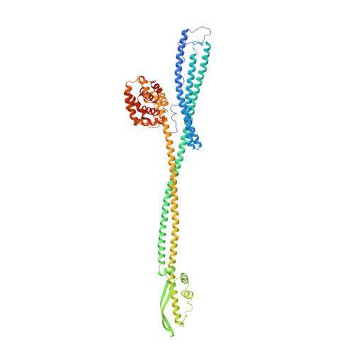
Crystal structure of colicin Ia.

Wiener, M.Freymann, D.Ghosh, P.Stroud, R.M.

(1997) Nature 385: 461-464

  • DOI: https://doi.org/10.1038/385461a0
  • Primary Citation of Related Structures:  
    1CII

  • PubMed Abstract: 

    The ion-channel forming colicins A, B, E1, Ia, Ib and N all kill bacterial cells selectively by co-opting bacterial active-transport pathways and forming voltage-gated ion conducting channels across the plasma membrane of the target bacterium. The crystal structure of colicin Ia reveals a molecule 210 A long with three distinct functional domains arranged along a backbone of two extraordinarily long alpha-helices. A central domain at the bend of the hairpin-like structure mediates specific recognition and binding to an outer-membrane receptor. A second domain mediates translocation across the outer membrane via the TonB transport pathway; the TonB-box recognition element of colicin Ia is on one side of three 80 A-long helices arranged as a helical sheet. A third domain is made up of 10 alpha-helices which form a voltage-activated and voltage-gated ion conducting channel across the plasma membrane of the target cell. The two 160 A-long alpha-helices that link the receptor-binding domain to the other domains enable the colicin Ia molecule to span the periplasmic space and contact both the outer and plasma membranes simultaneously during function.


  • Organizational Affiliation

    S-964 Department of Biochemistry and Biophysics, University of California, San Francisco 94143-0448, USA.


Macromolecules
Find similar proteins by:  (by identity cutoff)  |  3D Structure
Entity ID: 1
MoleculeChains Sequence LengthOrganismDetailsImage
COLICIN IA602Escherichia coliMutation(s): 0 
Gene Names: CIA
UniProt
Find proteins for P06716 (Escherichia coli)
Explore P06716 
Go to UniProtKB:  P06716
Entity Groups  
Sequence Clusters30% Identity50% Identity70% Identity90% Identity95% Identity100% Identity
UniProt GroupP06716
Sequence Annotations
Expand
  • Reference Sequence
Experimental Data & Validation

Experimental Data

  • Method: X-RAY DIFFRACTION
  • Resolution: 3.00 Å
  • R-Value Free: 0.312 
  • R-Value Work: 0.241 
  • R-Value Observed: 0.241 
  • Space Group: C 2 2 21
Unit Cell:
Length ( Å )Angle ( ˚ )
a = 64.4α = 90
b = 178.6β = 90
c = 285.5γ = 90
Software Package:
Software NamePurpose
PHASESphasing
SOLOMONphasing
X-PLORmodel building
XTALVIEWrefinement
X-PLORrefinement
DENZOdata reduction
SCALEPACKdata scaling
X-PLORphasing

Structure Validation

View Full Validation Report


Entry History 

Deposition Data

Revision History  (Full details and data files)

  • Version 1.0: 1998-01-14
    Type: Initial release
  • Version 1.1: 2008-03-24
    Changes: Version format compliance
  • Version 1.2: 2011-07-13
    Changes: Version format compliance
  • Version 1.3: 2024-02-07
    Changes: Data collection, Database references