1CI7

TERNARY COMPLEX OF THYMIDYLATE SYNTHASE FROM PNEUMOCYSTIS CARINII


Experimental Data Snapshot

  • Method: X-RAY DIFFRACTION
  • Resolution: 2.60 Å
  • R-Value Free: 0.294 
  • R-Value Work: 0.221 
  • R-Value Observed: 0.221 

wwPDB Validation   3D Report Full Report


This is version 1.4 of the entry. See complete history


Literature

The structural mechanism for half-the-sites reactivity in an enzyme, thymidylate synthase, involves a relay of changes between subunits.

Anderson, A.C.O'Neil, R.H.DeLano, W.L.Stroud, R.M.

(1999) Biochemistry 38: 13829-13836

  • DOI: https://doi.org/10.1021/bi991610i
  • Primary Citation of Related Structures:  
    1CI7

  • PubMed Abstract: 

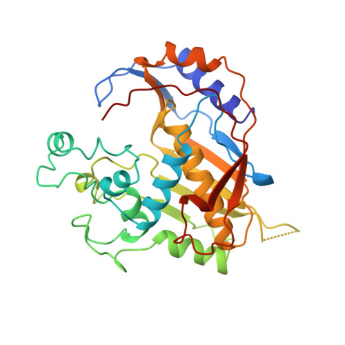
    Thymidylate synthase (TS), a half-the-sites reactive enzyme, catalyzes the final step in the de novo biosynthesis of deoxythymidine monophosphate, dTMP, required for DNA replication. The cocrystal structure of TS from Pneumocystis carinii (PcTS), a new drug target for an important pathogen, with its substrate, deoxyuridine monophosphate (dUMP), and a cofactor mimic, CB3717, was determined. The structure, solved at 2.6 A resolution, shows an asymmetric dimer with two molecules of the substrate dUMP bound yet only one molecule of cofactor analogue bound. The structural evidence reveals that upon binding cofactor analogue and forming a covalent bond from the nucleophilic cysteine to the substrate, dUMP, at one active site, PcTS undergoes a conformational change that renders the opposite monomer incapable of forming a covalent bond or binding a molecule of cofactor analogue. The communication pathway between the two active sites is evident, allowing a structural definition of the basis of half-the-sites reactivity for thymidylate synthase and providing an example of such a mechanism for other half-the-sites reactive enzymes.


  • Organizational Affiliation

    Macromolecular Structure Group, Department of Biochemistry and Biophysics, University of California at San Francisco 94143-0448, USA.


Macromolecules
Find similar proteins by:  (by identity cutoff)  |  3D Structure
Entity ID: 1
MoleculeChains Sequence LengthOrganismDetailsImage
PROTEIN (THYMIDYLATE SYNTHASE)
A, B
297Pneumocystis cariniiMutation(s): 0 
EC: 2.1.1.45
UniProt
Find proteins for P13100 (Pneumocystis carinii)
Explore P13100 
Go to UniProtKB:  P13100
Entity Groups  
Sequence Clusters30% Identity50% Identity70% Identity90% Identity95% Identity100% Identity
UniProt GroupP13100
Sequence Annotations
Expand
  • Reference Sequence
Small Molecules
Ligands 2 Unique
IDChains Name / Formula / InChI Key2D Diagram3D Interactions
CB3
Query on CB3

Download Ideal Coordinates CCD File 
D [auth A]10-PROPARGYL-5,8-DIDEAZAFOLIC ACID
C24 H23 N5 O6
LTKHPMDRMUCUEB-IBGZPJMESA-N
UMP
Query on UMP

Download Ideal Coordinates CCD File 
C [auth A],
E [auth B]
2'-DEOXYURIDINE 5'-MONOPHOSPHATE
C9 H13 N2 O8 P
JSRLJPSBLDHEIO-SHYZEUOFSA-N
Binding Affinity Annotations 
IDSourceBinding Affinity
CB3 PDBBind:  1CI7 Ki: 90 (nM) from 1 assay(s)
Experimental Data & Validation

Experimental Data

  • Method: X-RAY DIFFRACTION
  • Resolution: 2.60 Å
  • R-Value Free: 0.294 
  • R-Value Work: 0.221 
  • R-Value Observed: 0.221 
  • Space Group: P 21 21 21
Unit Cell:
Length ( Å )Angle ( ˚ )
a = 66.16α = 90
b = 178.76β = 90
c = 54.05γ = 90
Software Package:
Software NamePurpose
X-PLORmodel building
CNSrefinement
DENZOdata reduction
SCALEPACKdata scaling
X-PLORphasing

Structure Validation

View Full Validation Report


Entry History 

Deposition Data

Revision History  (Full details and data files)

  • Version 1.0: 2000-04-10
    Type: Initial release
  • Version 1.1: 2008-04-26
    Changes: Version format compliance
  • Version 1.2: 2011-07-13
    Changes: Version format compliance
  • Version 1.3: 2023-12-27
    Changes: Data collection, Database references, Derived calculations
  • Version 1.4: 2024-04-03
    Changes: Refinement description