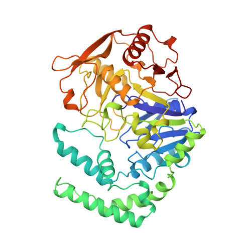
1CG4

STRUCTURE OF THE MUTANT (R303L) OF ADENYLOSUCCINATE SYNTHETASE FROM E. COLI COMPLEXED WITH, GDP, 6-PHOSPHORYL-IMP, AND MG2+


Experimental Data Snapshot

  • Method: X-RAY DIFFRACTION
  • Resolution: 2.50 Å
  • R-Value Free: 0.252 
  • R-Value Work: 0.179 

wwPDB Validation   3D Report Full Report


This is version 1.5 of the entry. See complete history


Literature

Mechanistic implications from crystalline complexes of wild-type and mutant adenylosuccinate synthetases from Escherichia coli.

Choe, J.Y.Poland, B.W.Fromm, H.J.Honzatko, R.B.

(1999) Biochemistry 38: 6953-6961

  • DOI: https://doi.org/10.1021/bi990159s
  • Primary Citation of Related Structures:  
    1CG0, 1CG1, 1CG3, 1CG4

  • PubMed Abstract: 

    Asp13 and His41 are essential residues of adenylosuccinate synthetase, putatively catalyzing the formation of adenylosuccinate from an intermediate of 6-phosphoryl-IMP. Wild-type adenylosuccinate synthetase and three mutant synthetases (Arg143 --> Leu, Lys16 --> Gln, and Arg303 --> Leu) from Eschericha coli have been crystallized in the presence of IMP, hadacidin (an analogue of L-aspartate), Mg2+, and GTP. The active site of each complex contains 6-phosphoryl-IMP, Mg2+, GDP, and hadacidin, except for the Arg303 --> Leu mutant, which does not bind hadacidin. In response to the formation of 6-phosphoryl-IMP, Asp13 enters the inner coordination sphere of the active site Mg2+. His41 hydrogen bonds with 6-phosphoryl-IMP, except in the Arg303 --> Leu complex, where it remains bound to the guanine nucleotide. Hence, recognition of the active site Mg2+ by Asp13 evidently occurs after the formation of 6-phosphoryl-IMP, but recognition of the intermediate by His41 may require the association of L-aspartate with the active site. Structures reported here support a mechanism in which Asp13 and His41 act as the catalytic base and acid, respectively, in the formation of 6-phosphoryl-IMP, and then act together as catalytic acids in the subsequent formation of adenylosuccinate.


  • Organizational Affiliation

    Department of Biochemistry, Biophysics, and Molecular Biology, Iowa State University, Ames 50011, USA.


Macromolecules
Find similar proteins by:  (by identity cutoff)  |  3D Structure
Entity ID: 1
MoleculeChains Sequence LengthOrganismDetailsImage
PROTEIN (ADENYLOSUCCINATE SYNTHETASE)431Escherichia coli K-12Mutation(s): 1 
EC: 6.3.4.4
UniProt
Find proteins for P0A7D4 (Escherichia coli (strain K12))
Explore P0A7D4 
Go to UniProtKB:  P0A7D4
Entity Groups  
Sequence Clusters30% Identity50% Identity70% Identity90% Identity95% Identity100% Identity
UniProt GroupP0A7D4
Sequence Annotations
Expand
  • Reference Sequence
Small Molecules
Experimental Data & Validation

Experimental Data

  • Method: X-RAY DIFFRACTION
  • Resolution: 2.50 Å
  • R-Value Free: 0.252 
  • R-Value Work: 0.179 
  • Space Group: P 32 2 1
Unit Cell:
Length ( Å )Angle ( ˚ )
a = 80.21α = 90
b = 80.21β = 90
c = 157.87γ = 120
Software Package:
Software NamePurpose
X-PLORmodel building
X-PLORrefinement
X-GENdata reduction
X-GENdata scaling
X-PLORphasing

Structure Validation

View Full Validation Report


Entry History 

Deposition Data

Revision History  (Full details and data files)

  • Version 1.0: 1999-06-17
    Type: Initial release
  • Version 1.1: 2008-04-26
    Changes: Version format compliance
  • Version 1.2: 2011-07-13
    Changes: Derived calculations, Version format compliance
  • Version 1.3: 2017-10-04
    Changes: Refinement description
  • Version 1.4: 2018-03-14
    Changes: Database references
  • Version 1.5: 2023-08-09
    Changes: Data collection, Database references, Derived calculations, Refinement description