1CDL

TARGET ENZYME RECOGNITION BY CALMODULIN: 2.4 ANGSTROMS STRUCTURE OF A CALMODULIN-PEPTIDE COMPLEX


Experimental Data Snapshot

  • Method: X-RAY DIFFRACTION
  • Resolution: 2.00 Å
  • R-Value Work: 0.208 
  • R-Value Observed: 0.208 

wwPDB Validation   3D Report Full Report


This is version 1.3 of the entry. See complete history


Literature

Target enzyme recognition by calmodulin: 2.4 A structure of a calmodulin-peptide complex.

Meador, W.E.Means, A.R.Quiocho, F.A.

(1992) Science 257: 1251-1255

  • DOI: https://doi.org/10.1126/science.1519061
  • Primary Citation of Related Structures:  
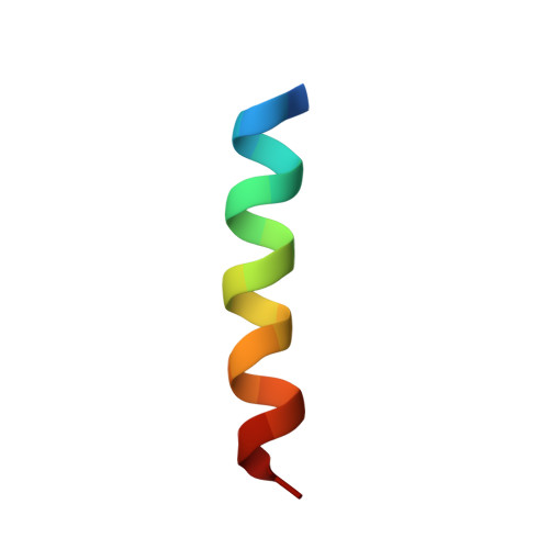
    1CDL

  • PubMed Abstract: 

    The crystal structure of calcium-bound calmodulin (Ca(2+)-CaM) bound to a peptide analog of the CaM-binding region of chicken smooth muscle myosin light chain kinase has been determined and refined to a resolution of 2.4 angstroms (A). The structure is compact and has the shape of an ellipsoid (axial ratio approximately 2:1). The bound CaM forms a tunnel diagonal to its long axis that engulfs the helical peptide, with the hydrophobic regions of CaM melded into a single area that closely covers the hydrophobic side of the peptide. There is a remarkably high pseudo-twofold symmetry between the closely associated domains. The central helix of the native CaM is unwound and expanded into a bend between residues 73 and 77. About 185 contacts (less than 4 A) are formed between CaM and the peptide, with van der Waals contacts comprising approximately 80% of this total.


  • Organizational Affiliation

    Howard Hughes Medical Institute, Baylor College of Medicine, Houston, TX 77030.


Macromolecules
Find similar proteins by:  (by identity cutoff)  |  3D Structure
Entity ID: 1
MoleculeChains Sequence LengthOrganismDetailsImage
CALMODULINA,
C [auth B],
E [auth C],
G [auth D]
147Homo sapiensMutation(s): 0 
UniProt & NIH Common Fund Data Resources
Find proteins for P0DP23 (Homo sapiens)
Explore P0DP23 
Go to UniProtKB:  P0DP23
PHAROS:  P0DP23
GTEx:  ENSG00000198668 
Entity Groups  
Sequence Clusters30% Identity50% Identity70% Identity90% Identity95% Identity100% Identity
UniProt GroupP0DP23
Sequence Annotations
Expand
  • Reference Sequence

Find similar proteins by:  Sequence   |   3D Structure  

Entity ID: 2
MoleculeChains Sequence LengthOrganismDetailsImage
CALCIUM/CALMODULIN-DEPENDENT PROTEIN KINASE TYPE II ALPHA CHAINB [auth E],
D [auth F],
F [auth G],
H
20Gallus gallusMutation(s): 0 
Gene Names: Mylk
UniProt
Find proteins for P11799 (Gallus gallus)
Explore P11799 
Go to UniProtKB:  P11799
Entity Groups  
Sequence Clusters30% Identity50% Identity70% Identity90% Identity95% Identity100% Identity
UniProt GroupP11799
Sequence Annotations
Expand
  • Reference Sequence
Small Molecules
Ligands 1 Unique
IDChains Name / Formula / InChI Key2D Diagram3D Interactions
CA
Query on CA

Download Ideal Coordinates CCD File 
I [auth A]
J [auth A]
K [auth A]
L [auth A]
M [auth B]
I [auth A],
J [auth A],
K [auth A],
L [auth A],
M [auth B],
N [auth B],
O [auth B],
P [auth B],
Q [auth C],
R [auth C],
S [auth C],
T [auth C],
U [auth D],
V [auth D],
W [auth D],
X [auth D]
CALCIUM ION
Ca
BHPQYMZQTOCNFJ-UHFFFAOYSA-N
Experimental Data & Validation

Experimental Data

  • Method: X-RAY DIFFRACTION
  • Resolution: 2.00 Å
  • R-Value Work: 0.208 
  • R-Value Observed: 0.208 
  • Space Group: P 21 21 2
Unit Cell:
Length ( Å )Angle ( ˚ )
a = 166.1α = 90
b = 104.9β = 90
c = 43.32γ = 90
Software Package:
Software NamePurpose
X-PLORmodel building
X-PLORrefinement
X-PLORphasing

Structure Validation

View Full Validation Report


Entry History 

Deposition Data

Revision History  (Full details and data files)

  • Version 1.0: 1994-08-31
    Type: Initial release
  • Version 1.1: 2008-03-03
    Changes: Version format compliance
  • Version 1.2: 2011-07-13
    Changes: Version format compliance
  • Version 1.3: 2024-02-07
    Changes: Data collection, Database references, Derived calculations, Other