1CCZ

CRYSTAL STRUCTURE OF THE CD2-BINDING DOMAIN OF CD58 (LYMPHOCYTE FUNCTION-ASSOCIATED ANTIGEN 3) AT 1.8-A RESOLUTION


Experimental Data Snapshot

  • Method: X-RAY DIFFRACTION
  • Resolution: 1.80 Å
  • R-Value Free: 0.243 
  • R-Value Work: 0.202 

wwPDB Validation   3D Report Full Report


This is version 1.4 of the entry. See complete history


Literature

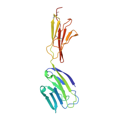
Crystal structure of the CD2-binding domain of CD58 (lymphocyte function-associated antigen 3) at 1.8-A resolution.

Ikemizu, S.Sparks, L.M.van der Merwe, P.A.Harlos, K.Stuart, D.I.Jones, E.Y.Davis, S.J.

(1999) Proc Natl Acad Sci U S A 96: 4289-4294

  • DOI: https://doi.org/10.1073/pnas.96.8.4289
  • Primary Citation of Related Structures:  
    1CCZ

  • PubMed Abstract: 

    The binding of the cell surface molecule CD58 (formerly lymphocyte function-associated antigen 3) to its ligand, CD2, significantly increases the sensitivity of antigen recognition by T cells. This was the first heterophilic cell adhesion interaction to be discovered and is now an important paradigm for analyzing the structural basis of cell-cell recognition. The crystal structure of a CD2-binding chimeric form of CD58, solved to 1.8-A resolution, reveals that the ligand binding domain of CD58 has the expected Ig superfamily V-set topology and shares several of the hitherto unique structural features of CD2, consistent with previous speculation that the genes encoding these molecules arose via duplication of a common precursor. Nevertheless, evidence for considerable divergence of CD2 and CD58 is also implicit in the structures. Mutations that disrupt CD2 binding map to the highly acidic surface of the AGFCC'C" beta-sheet of CD58, which, unexpectedly, lacks marked shape complementarity to the equivalent, rather more basic CD58-binding face of human CD2. The specificity of the very weak interactions of proteins mediating cell-cell recognition may often derive largely from electrostatic complementarity, with shape matching at the protein-protein interface being less exact than for interactions that combine specificity with high affinity, such as those involving antibodies.


  • Organizational Affiliation

    Laboratory of Molecular Biophysics, the Rex Richards Building, South Parks Road, Oxford, OX1 3QU, United Kingdom.


Macromolecules
Find similar proteins by:  (by identity cutoff)  |  3D Structure
Entity ID: 1
MoleculeChains Sequence LengthOrganismDetailsImage
PROTEIN (CD58)171Homo sapiensMutation(s): 0 
UniProt & NIH Common Fund Data Resources
Find proteins for P08921 (Rattus norvegicus)
Explore P08921 
Go to UniProtKB:  P08921
Find proteins for P19256 (Homo sapiens)
Explore P19256 
Go to UniProtKB:  P19256
PHAROS:  P19256
GTEx:  ENSG00000116815 
Entity Groups  
Sequence Clusters30% Identity50% Identity70% Identity90% Identity95% Identity100% Identity
UniProt GroupsP08921P19256
Sequence Annotations
Expand
  • Reference Sequence
Small Molecules
Ligands 1 Unique
IDChains Name / Formula / InChI Key2D Diagram3D Interactions
NAG
Query on NAG

Download Ideal Coordinates CCD File 
B [auth A],
C [auth A],
D [auth A]
2-acetamido-2-deoxy-beta-D-glucopyranose
C8 H15 N O6
OVRNDRQMDRJTHS-FMDGEEDCSA-N
Experimental Data & Validation

Experimental Data

  • Method: X-RAY DIFFRACTION
  • Resolution: 1.80 Å
  • R-Value Free: 0.243 
  • R-Value Work: 0.202 
  • Space Group: P 32 2 1
Unit Cell:
Length ( Å )Angle ( ˚ )
a = 116.43α = 90
b = 116.43β = 90
c = 51.52γ = 120
Software Package:
Software NamePurpose
DENZOdata reduction
SCALEPACKdata scaling
AMoREphasing
X-PLORrefinement

Structure Validation

View Full Validation Report


Entry History 

Deposition Data

Revision History  (Full details and data files)

  • Version 1.0: 1999-04-05
    Type: Initial release
  • Version 1.1: 2008-04-26
    Changes: Version format compliance
  • Version 1.2: 2011-07-13
    Changes: Non-polymer description, Version format compliance
  • Version 1.3: 2020-07-29
    Type: Remediation
    Reason: Carbohydrate remediation
    Changes: Data collection, Derived calculations, Structure summary
  • Version 1.4: 2023-08-09
    Changes: Data collection, Database references, Refinement description, Structure summary