Three-dimensional structure of adenosylcobinamide kinase/adenosylcobinamide phosphate guanylyltransferase from Salmonella typhimurium determined to 2.3 A resolution,.
Thompson, T.B., Thomas, M.G., Escalante-Semerena, J.C., Rayment, I.(1998) Biochemistry 37: 7686-7695
- PubMed: 9601028
- DOI: https://doi.org/10.1021/bi973178f
- Primary Citation of Related Structures:
1CBU - PubMed Abstract:
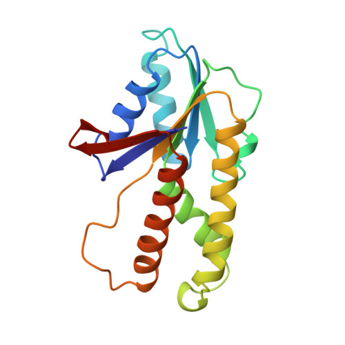
The X-ray structure of adenosylcobinamide kinase/adenosylcobinamide phosphate guanylyltransferase (CobU) from Salmonella typhimurium has been determined to 2.3 A resolution. This enzyme of subunit molecular weight 19 770 plays a central role in the assembly of the nucleotide loop for adenosylcobalamin where it catalyzes both the phosphorylation of the 1-amino-2-propanol side chain of the corrin ring and the subsequent attachment of GMP to form the product adenosylcobinamide-GDP. The kinase activity is believed to be associated with a P-loop motif, whereas the transferase activity proceeds at a different site on the enzyme via a guanylyl intermediate. The enzyme was crystallized in the space group C2221 with unit cell dimensions of a = 96.4 A, b = 114.4 A, and c = 106.7 A, with three subunits per asymmetric unit. The structure reveals that the enzyme is a molecular trimer and appears somewhat like a propeller with overall molecular dimensions of approximately 64 A x 77 A x 131 A. Each subunit consists of a single domain that is dominated by a seven-stranded mixed beta-sheet flanked on either side by a total of five alpha-helices and one helical turn. Six of the seven beta-strands run parallel. The C-terminal strand lies at the edge of the sheet and runs antiparallel to the others. Interestingly, CobU displays a remarkable structural and topological similarity to the central domain of the RecA protein, although the reason for this observation is unclear. The structure contains a P-loop motif located at the base of a prominent cleft formed by the association of two subunits and is most likely the kinase active site. Each subunit of CobU contains a cis peptide bond between Glu80 and Cys81 where Glu80 faces the P-loop and might serve to coordinate the magnesium ion of the triphosphate substrate. Interestingly, His46, which is the putative site for guanylylation, lies approximately 21 A from the P-loop and is solvent-exposed. This suggests that the enzyme undergoes a conformational change when the substrates bind to bring these two active sites into closer proximity.
Organizational Affiliation:
Institute for Enzyme Research, University of Wisconsin, Madison 53705, USA.